
Page 132 - Algae Anatomy, Biochemistry, and Biotechnology
P. 132
Anatomy 115
envelope. When the chloroplasts are located close to the nucleus, the chloroplast endoplasmic
reticulum is continuous with the nuclear envelope. In the Xanthophyceae, this connection is not
the rule. Thylakoids are grouped into lamellae of three, which are two in some Raphidophyceae,
with varying degrees of coherence depending on the species. Thylakoids from adjacent lamellae fre-
quently interconnect across the stroma. In all the classes, with the exception of Eustigmatophyceae
and Chattonella (Raphidophyceae), one lamella runs around the periphery of the chloroplast beneath
the chloroplast envelope, enclosing all the other lamellae. The lamella is called girdle lamella.
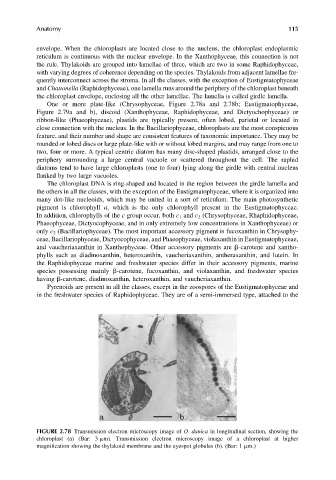
One or more plate-like (Chrysophyceae, Figure 2.78a and 2.78b; Eustigmatophyceae,
Figure 2.79a and b), discoid (Xanthophyceae, Raphidophyceae, and Dictyochophyceae) or
ribbon-like (Phaeophyceae), plastids are typically present, often lobed, parietal or located in
close connection with the nucleus. In the Bacillariophyceae, chloroplasts are the most conspicuous
feature, and their number and shape are consistent features of taxonomic importance. They may be
rounded or lobed discs or large plate-like with or without lobed margins, and may range from one to
two, four or more. A typical centric diatom has many disc-shaped plastids, arranged close to the
periphery surrounding a large central vacuole or scattered throughout the cell. The raphid
diatoms tend to have large chloroplasts (one to four) lying along the girdle with central nucleus
flanked by two large vacuoles.
The chloroplast DNA is ring-shaped and located in the region between the girdle lamella and
the others in all the classes, with the exception of the Eustigmatophyceae, where it is organized into
many dot-like nucleoids, which may be united in a sort of reticulum. The main photosynthetic
pigment is chlorophyll a, which is the only chlorophyll present in the Eustigmatophyceae.
In addition, chlorophylls of the c group occur, both c 1 and c 2 (Chrysophyceae, Rhaphidophyceae,
Phaeophyceae, Dictyocophyceae, and in only extremely low concentrations in Xanthophyceae) or
only c 2 (Bacillariophyceae). The most important accessory pigment is fucoxanthin in Chrysophy-
ceae, Bacillariophyceae, Dictyocophyceae, and Phaeophyceae, violaxanthin in Eustigmatophyceae,
and vaucheriaxanthin in Xanthophyceae. Other accessory pigments are b-carotene and xantho-
phylls such as diadinoxanthin, heteroxanthin, vaucheriaxanthin, antheraxanthin, and lutein. In
the Raphidophyceae marine and freshwater species differ in their accessory pigments, marine
species possessing mainly b-carotene, fucoxanthin, and violaxanthin, and freshwater species
having b-carotene, diadinoxanthin, heteroxanthin, and vaucheriaxanthin.
Pyrenoids are present in all the classes, except in the zoospores of the Eustigmatophyceae and
in the freshwater species of Raphidophyceae. They are of a semi-immersed type, attached to the
FIGURE 2.78 Transmission electron microscopy image of O. danica in longitudinal section, showing the
chloroplast (a) (Bar: 3 mm). Transmission electron microscopy image of a chloroplast at higher
magnification showing the thylakoid membrane and the eyespot globules (b). (Bar: 1 mm.)