
Page 146 - Algae Anatomy, Biochemistry, and Biotechnology
P. 146
Anatomy 129
Euglenophyta
Trichocysts are present only in some phagotrophic species, such as Entosiphon and Peranema tri-
chophorum. In Peranema ejectile organelles or mucocysts are located beneath the cell surface, often
subtending the pellicular articulations. In transverse sections they appear as hollow tubes of amor-
phous material, with low electron density, enclosed within a vesicle bounded by single membrane,
often found near the Golgi apparatus; or they show a content of greater density, and may subtend the
pellicle or protrude through it. Mucocysts subtending the pellicular articulations are ejected from
the cell through the grooves. They are characterized by three distinct regions: an inner tube
1.20 mm long and 1.18 mm wide with helical striations; an outer tube 0.77 mm long and
0.21 mm wide with diamond-shaped pattern; and a dense middle band 0.07 mm in width. Often
a crown of dense fibrillar material occurs at the tips of the mucocyst (Figure 2.86). Small slime-
extruding mucus bodies are the trichocyst counterparts commonly found in the non-phagotrophs.
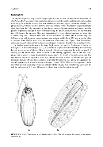
A feeding apparatus is present in many Euglenophyceae, such as Diplonema, Ploeotia, or
Entosiphon. In the latter (Figure 2.87a), it consists of a cytostome surrounded by four centrally
located vanes (series of plicate folds or curved ribbons) and supported by rods composed of
closely packed microtubules. Near the base of the feeding apparatus, one of the rods splits
giving rise to a total of three stout rods that extend nearly the length of the cell. About one third
the distance down the apparatus (from the anterior end) the number of microtubules per rod
increases dramatically and then decreases in number towards the base giving the apparatus the
overall appearance of a cone with one side open (Figure 2.87b). This feeding apparatus can be
moved in and out, extending toward the anterior of the cell and then withdrawing down into the
cell for a distance of 3–5 mm. This motion allows food to be drawn into the cell.
FIGURE 2.87 Cell of Entosiphon sp. (a); longitudinal section of the cell, showing the structure of the feeding
apparatus (b).