
Page 23 - Advances in Forensic Applications of Mass Spectrometry - Jehuda Yinon
P. 23
1522_C01.fm Page 10 Tuesday, December 2, 2003 10:05 AM
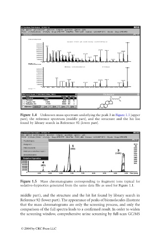
Figure 1.4 Unknown mass spectrum underlying the peak 3 in Figure 1.1 (upper
part), the reference spectrum (middle part), and the structure and the hit list
found by library search in Reference 92 (lower part).
Figure 1.5 Mass chromatograms corresponding to fragment ions typical for
sedative–hypnotics generated from the same data file as used for Figure 1.1.
middle part), and the structure and the hit list found by library search in
Reference 92 (lower part). The appearance of peaks of biomolecules illustrate
that the mass chromatograms are only the screening process, and only the
comparison of the full spectra leads to a confirmed result. In order to widen
the screening window, comprehensive urine screening by full-scan GC/MS
© 2004 by CRC Press LLC