
Page 42 - Advances in Forensic Applications of Mass Spectrometry - Jehuda Yinon
P. 42
1522_C01.fm Page 29 Tuesday, December 2, 2003 10:05 AM
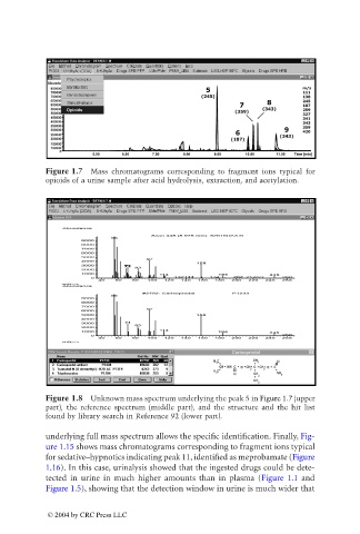
Figure 1.7 Mass chromatograms corresponding to fragment ions typical for
opioids of a urine sample after acid hydrolysis, extraction, and acetylation.
Figure 1.8 Unknown mass spectrum underlying the peak 5 in Figure 1.7 (upper
part), the reference spectrum (middle part), and the structure and the hit list
found by library search in Reference 92 (lower part).
underlying full mass spectrum allows the specific identification. Finally, Fig-
ure 1.15 shows mass chromatograms corresponding to fragment ions typical
for sedative–hypnotics indicating peak 11, identified as meprobamate (Figure
1.16). In this case, urinalysis showed that the ingested drugs could be dete-
tected in urine in much higher amounts than in plasma (Figure 1.1 and
Figure 1.5), showing that the detection window in urine is much wider that
© 2004 by CRC Press LLC