
Page 139 - Algae
P. 139
122 Algae: Anatomy, Biochemistry, and Biotechnology
nucleus may be less than 2 mm in diameter. The chromosomes retain their condensed condition
throughout interphase, appearing as granular or filamentous threads (Figure 2.81a and 2.81b). In
some nuclei the chromosomes radiate from the central endosome, while in others (even in the
same species) they coil haphazardly throughout the nucleoplasm. Mitosis begins with a forward
migration of the nucleus so that it comes to lie immediately posterior to the reservoir. In species
with several endosomes in the interphase nucleus, these usually fuse to form a single body. The
endosome then elongates along the division axis, perpendicular to the long axis of the cell, and
the chromosomes orient into the metaphase position, following three main types of orientation:
. Pairs of chromatids from late prophase orient into a circlet of single chromatids, separation
and segregation having occurred during orientation (E. gracilis)
. Pairs of chromatids from interphase or prophase come to lie along the division axis still as
pairs, parallel to one another and to the elongated endosomes (Euglena communis, Euglena
viridis)
. Single chromosomes from prophase line up along the division axis and there undergo dupli-
cation into the pairs of chromatids of that mitosis (E. acus, Euglena spirogyra)
These different types overlap to a certain extent, species differ mainly in the time at which the
double structure of the chromosomes first becomes microscopically visible. In all cases, the endo-
some continues to elongate and the chromatids segregate towards the ends of the endosome into two
daughter groups. This stage, with most but not quite all of the daughter chromosomes separated,
must be called metaphase in Euglena. During this early-to-late metaphase succession, the loco-
motor apparatus (flagella, photoreceptor, and eyespot) replicates and the reservoir divides. The
daughter reservoirs open into the still single canal, but each now has its own contractile vacuole,
eyespot, and flagella. Separation, segregation, and anaphasic movements of the chromatids are irre-
gular, and this, coupled with a very low chromatid velocity, results in an extremely long anaphase.
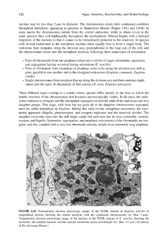
FIGURE 2.81 Transmission electron microscopy image of the WZSL mutant of Euglena gracilis in
longitudinal section, showing the central nucleous with the condensed chromosomes (a) (Bar: 3 mm).
Transmission electron microscopy image of the nucleus of the WZSL mutant of E. gracilis, showing the
nucleolus, the satellite nucleoli and the nuclear membrane pores (arrowhead) (b). (Bar: 0.3 mm.) (Courtesy
of Dr. Giovanna Rosati.)