
Page 33 - Algae
P. 33
16 Algae: Anatomy, Biochemistry, and Biotechnology
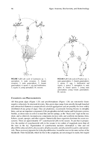
FIGURE 1.23 Life cycle of Laminaria sp.: 1, FIGURE 1.24 Life cycle of Porphyra sp.: 1,
0
0
sporophyte; 2, male zoospore; 2 , female male gametophyte; 1 , female gametophyte;
0
0
zoospore; 3, male gametophyte; 3 , female 2, sperm; 2 , egg; 3, fertilization and
0
gametophyte; 4, sperm; 4 , egg and fertilization; zygote; 4, spores; 5, sporophyte; 6, male
0
5, zygote; 6, young sporophyte. R!, meiosis. spore; 6 , female spores; 7, young male
gametophyte; young female gametophyte.
R!, meiosis.
CYANOPHYTA AND PROCHLOROPHYTA
All blue-green algae (Figure 1.25) and prochlorophytes (Figure 1.26) are non-motile Gram-
negative eubacteria. In structural diversity, blue-green algae range from unicells through branched
and unbranched filaments to unspecialized colonial aggregations and are possibly the most widely
distributed of any group of algae. They are planktonic, occasionally forming blooms in eutrophic
lakes, and are an important component of the picoplankton in both marine and freshwater systems;
benthic, as dense mats on soil or in mud flats and hot springs, as the “black zone” high on the sea-
shore, and as relatively inconspicuous components in most soils; and symbiotic in diatoms, ferns,
lichens, cycads, sponges, and other systems. Numerically these organisms dominate the ocean eco-
systems. There are approximately 10 24 cyanobacterial cells in the oceans. To put that in perspec-
tive, the number of cyanobacterial cells in the oceans is two orders of magnitude more than all
the stars in the sky. Pigmentation of cyanobacteria includes chlorophyll a, blue and red
phycobilins (phycoerythrin, phycocyanin, allophycocyanin, and phycoerythrocyanin), and caroten-
oids. These accessory pigments lie in the phycobilisomes, located in rows on the outer surface of the
thylakoids. Their thylakoids, which lie free in the cytoplasm, are not arranged in stacks, but singled