
Page 75 - Algae
P. 75
58 Algae: Anatomy, Biochemistry, and Biotechnology
algae such as Chlorophyceae and Charopyceae. Within this group, there are few genera whose fla-
gella differ in length, which are termed “anisokont.”
Description of flagella anatomy will proceed from outside to the inside, from the surface
features and components to the axoneme and additional inclusions to the structures anchoring
the flagella to the cell.
Flagellar Shape and Surface Features
Deviations from the cylindrical shape are rare among the algae. Usually the flagellar membrane fits
smoothly around the axoneme and a total diameter of 0.25–0.35 mm, excluding scales, hairs, etc.,
holds for most species. If extra material is present between the axoneme and the flagellar mem-
brane, the flagellum diameter increases either locally as in the case of flagellar swellings, or
through almost the entire length as in the case of paraxial rods. Minor deviations from the cylind-
rical shape are caused by small extensions of the membrane to form one or more longitudinal keels
running the length of the flagellum. Greater extension of the membrane forms a ribbon or wing sup-
ported along the edge by a paraxial rod. More variations are present in the flagellar tip, because
flagella can possess a hairpoint, that is, their distal part is thinner with respect to the rest of the fla-
gellum or blunt-tipped, with an abundance of intermediates between these two types.
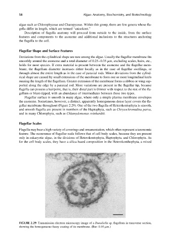
Flagellar surface is smooth in many algae, where only a simple plasma membrane envelopes
the axoneme. Sometimes, however, a distinct, apparently homogeneous dense layer covers the fla-
gellar membrane throughout (Figure 2.29). One of the two flagella of Heterokontophyta is smooth,
and smooth flagella are present in members of the Haptophyta, such as Chrysochromulina parva,
and in many Chlorophyta, such as Chlamydomonas reinhardtii.
Flagellar Scales
Flagella may bear a high variety of coverings and ornamentation, which often represent a taxonomic
feature. The occurrence of flagellar scale follows that of cell body scales, because they are present
only in eukaryotic algae, in the divisions of Heterokontophyta, Haptophyta, and Chlorophyta. As
for the cell body scales, they have a silica-based composition in the Heterokonthophyta, a mixed
FIGURE 2.29 Transmission electron microscopy image of a Dunaliella sp. flagellum in transverse section,
showing the homogeneous fuzzy coating of its membrane. (Bar: 0.10 mm.)