
Page 237 - Bio Engineering Approaches to Cancer Diagnosis and Treatment
P. 237
236 CHAPTER 9 Application of microfluidics in cancer treatment
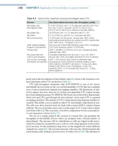
Table 9.3 Some of the important existing technologies about CTC.
Devices Key observations (recovery rate, throughput, purity)
Micropillar chip; 5-1281 CTCs/mL with 1 mL/h, detection rate 65%; 63 CTCs/
Herringebone chip mL with 1.2 mL/h, detection rate 91.8 ± 5% for PC3;
50.3 CTCs/mL with 8 mL/h, detection rate 98% SKBR3;
Micropillar chip 30 CTCs/mL with 1 mL/h, detection rate 97 ± 3%
Microfilter 0–12.5 CTCs/mL with 90 mL/h, detection rate 90%
Microchannel array 10 CTCs/mL with 36 µL/min, recover rate >95%. Using
additional grooved surface can achieve over 90% capture rate
with >84% purity. Throughput: 3.6 mL/h.
µHall: antibody labeled Recovery rate of MDA-MB-468 99%; purity 100%; throughput
magnetic nanoparticles 3.25 mL/h; analyzing ovarian 7.6 CTCs/mL.
Portable filter MCF-7, SK-BR-3: 5 CTCs/mL at 0.5 psi, constant pressure,
detection rate >90%
Microsieve filter with Undiluted whole blood with flow rate < 2 mL/min. MCF-7
uniform pore structure recover rate >80%; accurately detected CTC from 8 patients.
Lab-on-disc: centrifugal MCF-7: 61% capture rate; have the potential to high-
microfluidic size selection throughput and easy detection by directly reading on disc.
After collection, the MCF-7 still viable in 15 days cell culture.
Detect 0–90 CTCs/7.5 mL. Throughput: >3 mL/min;
sensitivity: 95.9 ± 3.1% recovery rate; selectivity: >2.5log
WBCs depletion.
purity lead to the development of microfluidic chip [62]. Some of the important exist-
ing technologies about CTC are listed in Table 9.3.
CTC high-throughput entrapment chip (CTC-HTECH) is one of the newest
microfluidic devices based on the size and deformability of CTC that have multiple
rows of microconstriction channels and trapping chambers. The limitations of inef-
ficient capture that were observed in existing size-dependent CTC capture designs
have been eliminated using CTC-HTECH. The blood cells in blood flow that includes
blood cells and CTCs pass through the microchannel, while single CTC is captured
in trapping chambers. The number of CTCs is determined when trapping chambers is
scanned. This ability is not available in other CTC microfluidic chips based on size.
The cells were then detached from the flask with a trypsin-EDTA solution (Sigma
Aldrich). The size of prostate cancer cells is in the range of 10–15 µm which is larger
than blood cells [62]. The schematic of the flow configuration for the acoustophore-
sis cell separation is shown in Fig. 9.9.
The use of a syringe pump to the creation of constant flow can guarantee the
throughput of microfluidic devices which are designed with a limited number of
microchannel. The pressure will be redistributed as cells are trapped in the micro-
channel by using the constant flow rate to a device with a large quantity of microchan-
nel in an array. So the mechanical drag forces on the trapped cells within different
channels are varied [62]. The increased pressure will cause the cell deformation that
could damage cells, leading to poor recovery of viable cells [69,70]. The alteration in