
Page 299 - Biodegradable Polyesters
P. 299
11.2 Simply Blended Drug/Biopolymer Nanofibers 277
can be continuously and gently fed through the spinneret. Under a high volt-
age, an evenly distributed electric field is formed and the liquid surface at the
end of the needle is charged. Once the electric field intensity exceeds the liquid
surface tension, the continuous jet flow of charged solution undergoes stretching
and elongation, in which process the solvent evaporates and the diameter of the
jet is significantly reduced to hundreds of nanometers to form nanofibers. There-
fore, drug-loaded single-component and multicomponent biopolymer nanofibers
can be easily obtained by electrospinning of the precursor drug/polymer blending
solution.
11.2.1
Drug-Loaded Single-Component Biopolymer Nanofibers
Natural biopolymers produced from various natural resources such as starch,
cellulose, and protein have been extremely popular for their abundant, renewable,
inexpensive, environmentally friendly, and biodegradable properties. Cellulose
acetate (CA), the acetate ester of cellulose, is the primary structural component
of the cell wall of green plants and one of the most common biopolymers on
earth. The fabrication of curcumin-loaded ultrafine CA fiber mats from neat CA
solution containing curcumin in various amounts was carried out by electro-
spinning, which was proven nontoxic to normal human dermal fibroblasts [16].
Electrospun CA nanofiber membranes were also used as carriers for delivery of
model vitamins, vitamin A acid and vitamin E, which exhibited a gradual and
monotonous increase in the cumulative release of the vitamins over the test
periods compared to a burst release of the vitamins from their corresponding
as-cast films [17]. Despite many advances in modern medicine, human immun-
odeficiency virus (HIV) still affects the health of millions of people worldwide.
Therefore, the potential use of electrospun cellulose acetate phthalate (CAP)
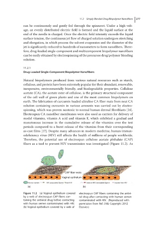
fibers as a tool to prevent HIV transmission was investigated (Figure 11.2). As
CAP fiber webs
Vaginal epithelium
Human semen HIV associated Sperm Free HIV Inactive HIV associated Sperm Inactive free HIV
(a) (b)
Figure 11.2 (a) Vaginal epithelium covered electrospun CAP fibers containing the antivi-
by a web of electrospun CAP fibers con- ral drug after contacting with human semen
taining the antiviral drug before contacting contaminated with HIV. (Reproduced with
with human semen contaminated with HIV. permission from Ref. [18]; Copyright 2012
(b) Vaginal epithelium covered by a web of Elsevier.)