
Page 492 - Carrahers_Polymer_Chemistry,_Eighth_Edition
P. 492
Testing and Spectrometric Characterization of Polymers 455
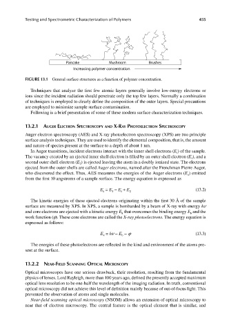
Pancake Mushroom Brushes
Increasing polymer concentration
FIGURE 13.1 General surface structures as a function of polymer concentration.
Techniques that analyze the first few atomic layers generally involve low-energy electrons or
ions since the incident radiation should penetrate only the top few layers. Normally a combination
of techniques is employed to clearly define the composition of the outer layers. Special precautions
are employed to minimize sample surface contamination.
Following is a brief presentation of some of these modern surface characterization techniques.
13.2.1 AUGER ELECTRON SPECTROSCOPY AND X-RAY PHOTOELECTRON SPECTROSCOPY
Auger electron spectroscopy (AES) and X-ray photoelectron spectroscopy (XPS) are two principle
surface analysis techniques. They are used to identify the elemental composition, that is, the amount
and nature of species present at the surface to a depth of about 1 nm.
In Auger transitions, incident electrons interact with the inner shell electrons (E ) of the sample.
i
The vacancy created by an ejected inner shell electron is filled by an outer shell electron (E ), and a
1
second outer shell electron (E ) is ejected leaving the atom in a doubly ionized state. The electrons
2
ejected from the outer shells are called Auger electrons, named after the Frenchman Pierre Auger,
who discovered the effect. Thus, AES measures the energies of the Auger electrons (E ) emitted
a
from the first 10 angstroms of a sample surface. The energy equation is expressed as
E = E − E + E 2 (13.2)
a
1
i
The kinetic energies of these ejected electrons originating within the first 30 Å of the sample
surface are measured by XPS. In XPS, a sample is bombarded by a beam of X-ray with energy hν
and core electrons are ejected with a kinetic energy E that overcomes the binding energy E and the
b
k
work function (ϕ). These core electrons are called the X-ray photoelectrons. The energy equation is
expressed as follows:
E = hν − E − ϕ (13.3)
b
k
The energies of these photoelectrons are reflected in the kind and environment of the atoms pre-
sent at the surface.
13.2.2 NEAR-FIELD SCANNING OPTICAL MICROSCOPY
Optical microscopes have one serious drawback, their resolution, resulting from the fundamental
physics of lenses. Lord Rayleigh, more than 100 years ago, defined the presently accepted maximum
optical lens resolution to be one-half the wavelength of the imaging radiation. In truth, conventional
optical microscopy did not achieve this level of definition mainly because of out-of-focus light. This
prevented the observation of atoms and single molecules.
Near-fi eld scanning optical microscopy (NSOM) allows an extension of optical microscopy to
near that of electron microscopy. The central feature is the optical element that is similar, and
9/14/2010 3:42:13 PM
K10478.indb 455 9/14/2010 3:42:13 PM
K10478.indb 455