
Page 189 - Computational Modeling in Biomedical Engineering and Medical Physics
P. 189
178 Computational Modeling in Biomedical Engineering and Medical Physics
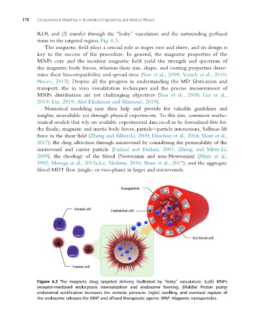
ROI, and (3) transfer through the “leaky” vasculature and the surrounding perfused
tissue to the targeted region, Fig. 6.3.
The magnetic field plays a crucial role at stages two and three, and its design is
key to the success of the procedure. In general, the magnetic properties of the
MNPs core and the incident magnetic field yield the strength and spectrum of
the magnetic body forces, whereas their size, shape, and coating properties deter-
mine their biocompatibility and spread time (Sun et al., 2008; Veiseh et al., 2010;
Nacev, 2013). Despite all the progress in understanding the MD fabrication and
transport, the in vivo visualization techniques and the precise measurement of
MNPs distribution are yet challenging objectives (Sun et al., 2008; Liu et al.,
2019; Liu, 2019; Abd Elrahman and Mansour, 2019).
Numerical modeling may then help and provide for valuable guidelines and
insights, unavailable yet through physical experiments. To this aim, consistent mathe-
matical models that rely on available experimental data need to be formulated first for:
the fluidic, magnetic and inertia body forces, particle particle interactions, Saffman lift
force in the shear field (Zheng and Silber-Li, 2009; Drochon et al., 2016; Shaw et al.,
2017), the drug advection through microvessel by considering the permeability of the
microvessel and carrier particle (Furlani and Furlani, 2007; Zheng and Silber-Li,
2009), the rheology of the blood (Newtonian and non-Newtonian) (Misra et al.,
1992; Morega et al., 2013a,b,c; S˘ av˘ astru, 2016; Shaw et al., 2017), and the aggregate
blood MDT flow (single- or two-phase) in larger and microvessels.
Figure 6.3 The magnetic drug targeted delivery facilitated by “leaky” vasculature. (Left) MNPs
receptor-mediated endocytosis internalization and endosome forming. (Middle) Proton pump
endosomal acidification increases the osmotic pressure; (right) swelling, and eventual rupture of
the endosome releases the MNP and affixed therapeutic agents. MNP, Magnetic nanoparticles.