
Page 104 -
P. 104
Section 3.1 Human Color Perception 72
1
0
S
1
M L
2
3
4
5
6
7
8
350 400 450 500 550 600 650 700 750 800 850
Wavelength in nm
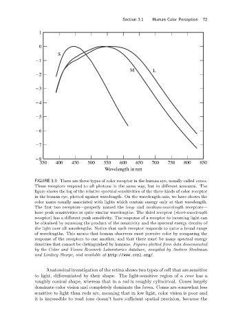
FIGURE 3.3: There are three types of color receptor in the human eye, usually called cones.
These receptors respond to all photons in the same way, but in different amounts. The
figure shows the log of the relative spectral sensitivities of the three kinds of color receptor
in the human eye, plotted against wavelength. On the wavelength axis, we have shown the
color name usually associated with lights which contain energy only at that wavelength.
The first two receptors—properly named the long- and medium-wavelength receptors—
have peak sensitivities at quite similar wavelengths. The third receptor (short-wavelength
receptor) has a different peak sensitivity. The response of a receptor to incoming light can
be obtained by summing the product of the sensitivity and the spectral energy density of
the light over all wavelengths. Notice that each receptor responds to quite a broad range
of wavelengths. This means that human observers must perceive color by comparing the
response of the receptors to one another, and that there must be many spectral energy
densities that cannot be distinguished by humans. Figures plotted from data disseminated
by the Color and Vision Research Laboratories database, compiled by Andrew Stockman
and Lindsey Sharpe, and available at http://www.cvrl.org/.
Anatomical investigation of the retina shows two types of cell that are sensitive
to light, differentiated by their shape. The light-sensitive region of a cone has a
roughly conical shape, whereas that in a rod is roughly cylindrical. Cones largely
dominate color vision and completely dominate the fovea. Cones are somewhat less
sensitive to light than rods are, meaning that in low light, color vision is poor and
it is impossible to read (one doesn’t have sufficient spatial precision, because the