
Page 95 - Electrical Safety of Low Voltage Systems
P. 95
78 Chapter Five
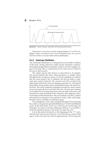
FIGURE 5.7 Burst of action potentials and resulting tetanization.
Tetanization occurs for currents ranging between 21 and 50 mA.
Higher values of current do not cause tetanization, but, as we see in
the next section, can cause other serious pathologies.
5.4.2 Ventricular Fibrillation
The ventricular fibrillation is a nonspontaneous reversible condition
of the heart, during which the cardiac muscle disorderly contracts,
denying the proper blood circulation, which is crucial to supply oxy-
gen to the body. This is considered to be the main cause of death in
the case of electrocution.
The cardiac muscle (also known as myocardium) is an involun-
tary muscle found in the heart, whose function is to “pump” blood
throughout the circulatory system. It has the capability to contract,
like any other muscles, but, in addition, also has the ability to gen-
erate and conduct electricity. The sinoatrial node (SA), located in the
right atrium of the heart, acts as an impulse generator (i.e., a biological
pacemaker) and generates action potentials that drive the heart con-
tractions. The action potentials propagate through the whole cardiac
muscle and reach the atrioventricular node (AV). As atria and ventricles
are insulated by nonconducting tissues, the AV node will receive and
transmit the action potentials to the ventricles, after applying a func-
tional delay to this transmission. The myocardium can, then, contract
and perform its important and continuous duties. After contracting,
the heart relaxes and fills up with blood again.
The propagation of the action potential through the heart during
the cardiac cycle generates potential differences V(t) between different
points of the entire body, which vary with time. By monitoring such
potentials, by means of electrocardiograms (EKGs), it is possible to
study the electrical activity of the heart over time (Fig. 5.8).
It is in this period of time dt, at the beginning of the “T” wave,
that the heart relaxes and awaits for a new stimulus to contract itself
again. In this time interval, which is approximately 150-ms long and