
Page 433 - Environmental Nanotechnology Applications and Impacts of Nanomaterials
P. 433
Toxicological Impacts of Nanomaterials 413
generally contains only Fe (III). It is in this form that most of the iron
in the body is stored (Dobson, 2001).
The reaction of the magnetic particles in a magnetic force has been
used in applications including drug targeting, bioseparation, and cell
sorting. Cell labeling with magnetic nanoparticles is an increasingly
common method for in vitro cell separation as well as for in vivo imag-
ing due to their signal amplification properties in magnetic resonance
imaging (MRI). Magnetic cell labeling is very promising for therapy, by
allowing for targeted magnetic intracellular hyperthermia (Ito et al.,
2001, 2005). All these applications require that cells efficiently capture
the magnetic nanoparticles either in vitro or in vivo. For in vitro stud-
ies, magnetic labeling only needs cellular uptake by the endocytosis
pathway, whereas in vivo, high affinity ligands needs to be grafted onto
nanoparticles surface for specific cellular interactions (Wilhelm et al.,
2003; Zhang et al., 2002). The primary problem encountered by all par-
ticles used in vivo is the adsorption of biological elements, especially pro-
teins (Portet et al., 2001; Ramge et al., 2000). Once the particles are
injected into the bloodstream, they are rapidly coated by plasma pro-
teins, a process known as opsonization, which is critical in dictating the
disposition of the injected particles (Davis et al., 1997). Normally,
opsonization renders the particles recognizable by the body’s major
defense system, the reticuloendothelial system (Araujo et al., 1999;
Berry et al., 2003; Kreuter et al., 1994).
The role of coating iron nanoparticles on the internalization efficiency
has been investigated in a series of studies by Wilhelm et al. (2003).
These authors compared cell uptake of anionic maghemite nanoparti-
cles (AMNP), which were coated with DMSA (meso-2,3-dimercaptosuc-
cinic acid), bovine serum albumin (BSA), or dextran. They quantified
particle uptake using new complementary magnetic assays, magne-
tophoresis, and electron spin resonance. After one hour of incubation in
mouse macrophages or human ovarian tumor HeLa cells with bare
AMNP, adhesion of the anionic nanoparticles on the plasma membrane
was seen mainly in the form of clusters. A few minutes later, densely con-
fined AMNP were located in various morphological forms within endo-
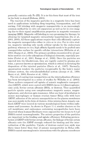
somes and lysosomes. As shown in Figure 11.4, similar clusters on the
cell membrane and endosomes containing nanoparticles have been
observed when human fibroblasts were exposed for two hours to 0.1 g/l
DMSA-coated nanomaghemite. The anionic properties of the particles
are important in the binding and uptake efficiency. Following preincu-
bation of AMNP with bovine serum albumin, the linkage of bovine serum
albumin onto the AMNP strongly reduced the binding and the inter-
nalization of the particles. Uptake of dextran-coated iron oxide was
three times lower than that of anionic nanoparticles in HeLa cells.
DMSA-coated nanomaghemite interactions with the plasma membrane