
Page 80 - Fundamentals of Light Microscopy and Electronic Imaging
P. 80
DEFINING DIFFRACTION AND INTERFERENCE 63
Aperture Specimen
θ 2
θ 1
(a) (b)
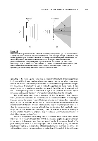
Figure 5-2
Diffraction at an aperture and at a substrate containing fine particles. (a) The electric field of
a planar wavefront becomes disturbed by diffraction upon passage through an aperture. The
waves appear to grab hold of the aperture and swing around into its geometric shadow. The
amplitude profile of a transmitted wavefront is also no longer uniform and remains
permanently altered after passage through the aperture (not shown). (b) A substrate
containing a layer of a mixture of fine particles (0.2 and 2 m diameter) diffracts an incident
planar wavefront into scattered beams that diverge at different angles. The angle of
spreading ( ) is inversely proportional to the size of the particles.
spreading of the beam depend on the size and density of the light-diffracting particles.
In the case of illuminated specimens in the microscope, there are therefore two primary
sites of diffraction: one at the specimen itself and another in the aperture of the objec-
tive lens. Image formation by a lens is critically dependent on these events. If light
passes through an object but does not become absorbed or diffracted, it remains invisi-
ble. It is the spreading action or diffraction of light at the specimen that allows objects
to become visible, and the theory of image formation is based on this principle.
Just as diffraction describes the scattering of light by an object into divergent
waves, interference describes the recombination and summation of two or more super-
imposed waves, the process responsible for creating the real intermediate image of an
object in the focal plane of a microscope. In a real sense, diffraction and interference are
manifestations of the same process. The traditional way of describing interference is to
show the recombination of waves graphically in a plot depicting their amplitude, wave-
length, and relative phase displacement (Fig. 5-3). The addition of two initial waves pro-
duces a resultant wave whose amplitude may be increased (constructive interference) or
diminished (destructive interference).
The term interference is frequently taken to mean that waves annihilate each other
if they are out of phase with each other by /2, and indeed a graphical depiction of inter-
fering waves reinforces this idea. However, since it can be demonstrated that all of the
photon energy delivered to a diffracting object such as a diffraction grating can be com-
pletely recovered (e.g., in a resultant diffraction pattern), it is clear that photons do not
self-annihilate; rather their energies become redistributed (channeled) during diffraction