
Page 381 - Handbook of Biomechatronics
P. 381
Current Advances in the Design of Retinal and Cortical Visual Prostheses 375
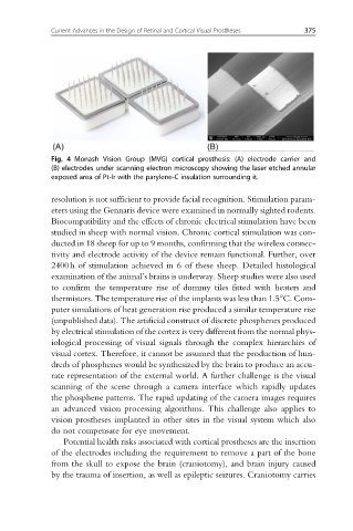
Fig. 4 Monash Vision Group (MVG) cortical prosthesis: (A) electrode carrier and
(B) electrodes under scanning electron microscopy showing the laser etched annular
exposed area of Pt-Ir with the parylene-C insulation surrounding it.
resolution is not sufficient to provide facial recognition. Stimulation param-
eters using the Gennaris device were examined in normally sighted rodents.
Biocompatibility and the effects of chronic electrical stimulation have been
studied in sheep with normal vision. Chronic cortical stimulation was con-
ducted in 18 sheep for up to 9 months, confirming that the wireless connec-
tivity and electrode activity of the device remain functional. Further, over
2400h of stimulation achieved in 6 of these sheep. Detailed histological
examination of the animal’s brains is underway. Sheep studies were also used
to confirm the temperature rise of dummy tiles fitted with heaters and
thermistors. The temperature rise of the implants was less than 1.5°C. Com-
puter simulations of heat generation rise produced a similar temperature rise
(unpublished data). The artificial construct of discrete phosphenes produced
by electrical stimulation of the cortex is very different from the normal phys-
iological processing of visual signals through the complex hierarchies of
visual cortex. Therefore, it cannot be assumed that the production of hun-
dreds of phosphenes would be synthesized by the brain to produce an accu-
rate representation of the external world. A further challenge is the visual
scanning of the scene through a camera interface which rapidly updates
the phosphene patterns. The rapid updating of the camera images requires
an advanced vision processing algorithms. This challenge also applies to
vision prostheses implanted in other sites in the visual system which also
do not compensate for eye movement.
Potential health risks associated with cortical prostheses are the insertion
of the electrodes including the requirement to remove a part of the bone
from the skull to expose the brain (craniotomy), and brain injury caused
by the trauma of insertion, as well as epileptic seizures. Craniotomy carries