
Page 171 - Inorganic Mass Spectrometry - Fundamentals and Applications
P. 171
Secondary Ion Mass Spectromet~
n
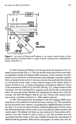
Ion source of Herzog and Viehbock: A, ion source (canal ray tube) at inde-
pendent potential; B, primary beam; C, sample; D and E, i~ersion lens, independent po-
tentials. (From Ref. 9.)
In 1962 Castaing and Slodzian [ 18,191 reported the development of the sec-
ondary ion microscope (Fig. 4.3). This was the first microscope to have mass analy-
In
sis capabilities and the first imaging SIMS instrument. this inst~ment, the sput-
tered ions are collected by an immersion lens, passed through a stigmatic magnetic
the
field, and projected onto an ion-to-electron converter that accelerates electrons
to a fluorescent screen, where a mass analyzed image of the sample surface is
formed. Rouberol et al., at Cameca Instruments, developed an improved version
of this instrument in 1968 [20,21], the IMS-300 (Fig. 4.4). Unique features of this
instrument were the Castaing-Henry magnetic prism [22,23] and an electrostatic
mirror for mass and energy filtration that maintained the optical axis of the mi-
croscope. The IMS-300 imaged a field of view 165-330 pm with a lateral resolu-
tion to about 1 pm Mass resolution of -1000 could be achieved.
In 1967 Liebl reported the development of the first imaging SIMS instru-
ment based on the principle of focused ion beam scanning [24]. This instrument,
the ion microprobe mass analyzer, was produced by Applied Research Laborato-
ries (Fig. 4.5). It used an improved hollow cathode duoplasmatron [25] ion source
in
that eliminated filaments used earlier sources and allowed stable operation with
reactive gases. The primary ion beam was mass analyzed for beam purity and fo-
cused in a two-lens column to a spot as small as 2 pm. The secondary ions were
accelerated from the sample surface into a double focusing mass spectrometer of
Mattauch-Herzog geometry. Both positive and negative secondary ions were de-