
Page 277 - Introduction to Paleobiology and The Fossil Record
P. 277
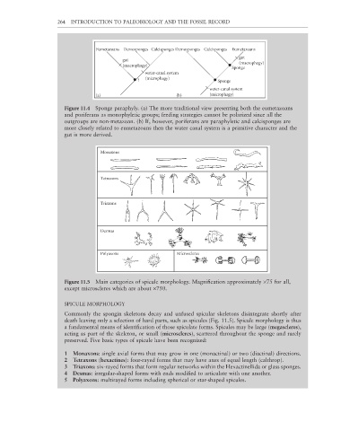
264  INTRODUCTION TO PALEOBIOLOGY AND THE FOSSIL RECORD


                                    Eumetazoans    Demosponges   Calcisponges Demosponges    Calcisponges     Eumetazoans
                                                                                        gut
                                              gut
                                                                                        (macrophagy)
                                              (macrophagy)
                                                                                     Sponge
                                                      water-canal system
                                                   ?  (microphagy)              Sponge
                                                                             water-canal system
                                    (a)                          (b)         (microphagy)

                        Figure 11.4  Sponge paraphyly. (a) The more traditional view presenting both the eumetazoans
                        and poriferans as monophyletic groups; feeding strategies cannot be polarized since all the
                        outgroups are non-metazoan. (b) If, however, poriferans are paraphyletic and calcisponges are
                        more closely related to eumetazoans then the water canal system is a primitive character and the
                        gut is more derived.


                                      Monaxons


                                      Tetraxons


                                      Triaxons


                                      Desmas


                                      Polyaxons                  Microscleres


                        Figure 11.5  Main categories of spicule morphology. Magnifi cation approximately ×75 for all,
                        except microscleres which are about ×750.

                        SPICULE MORPHOLOGY

                        Commonly the spongin skeletons decay and unfused spicular skeletons disintegrate shortly after
                        death leaving only a selection of hard parts, such as spicules (Fig. 11.5). Spicule morphology is thus

                        a fundamental means of identification of those spiculate forms. Spicules may be large (megascleres),
                        acting as part of the skeleton, or small (microscleres), scattered throughout the sponge and rarely
                        preserved. Five basic types of spicule have been recognized:

                        1  Monaxons: single axial forms that may grow in one (monactinal) or two (diactinal) directions.
                        2  Tetraxons (hexactines): four-rayed forms that may have axes of equal length (calthrop).
                        3  Triaxons: six-rayed forms that form regular networks within the Hexactinellida or glass sponges.
                        4  Desmas: irregular-shaped forms with ends modified to articulate with one another.

                        5  Polyaxons: multirayed forms including spherical or star-shaped spicules.