
Page 415 - Introduction to Paleobiology and The Fossil Record
P. 415
402 INTRODUCTION TO PALEOBIOLOGY AND THE FOSSIL RECORD
DORSAL
12 13 14 15
21
16
17
7
11 6
8
5
3
4 18
9 2
10
1
20
(a) 19
VENTRAL
Key
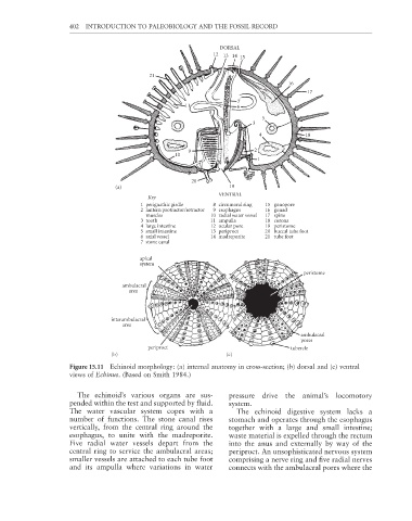
1 perignathic girdle 8 circumoral ring 15 gonopore
2 lantern protractor/retractor 9 esophagus 16 gonad
muscles 10 radial water vessel 17 spine
3 tooth 11 ampulla 18 corona
4 large intestine 12 ocular pore 19 peristome
5 small intestine 13 periproct 20 buccal tube foot
6 axial vessel 14 madreporite 21 tube foot
7 stone canal
apical
system
peristome
ambulacral
area
interambulacral
area
ambulacral
pores
periproct tubercle
(b) (c)
Figure 15.11 Echinoid morphology: (a) internal anatomy in cross-section; (b) dorsal and (c) ventral
views of Echinus. (Based on Smith 1984.)
The echinoid’s various organs are sus- pressure drive the animal’s locomotory
pended within the test and supported by fl uid. system.
The water vascular system copes with a The echinoid digestive system lacks a
number of functions. The stone canal rises stomach and operates through the esophagus
vertically, from the central ring around the together with a large and small intestine;
esophagus, to unite with the madreporite. waste material is expelled through the rectum
Five radial water vessels depart from the into the anus and externally by way of the
central ring to service the ambulacral areas; periproct. An unsophisticated nervous system
smaller vessels are attached to each tube foot comprising a nerve ring and fi ve radial nerves
and its ampulla where variations in water connects with the ambulacral pores where the