
Page 18 - Macromolecular Crystallography
P. 18
CLASSICAL CLONING, EXPRESSION, AND PURIFICATION 7
by immobilization on IgG (Nilsson et al., 1985). S-transferase as the fusion tag (Smith and John-
However, the drawback of using immunoaffin- son, 1988), which uses immobilized glutathione
ity procedures is that immunological detection can for isolation (Protocol 1.4). Both are commer-
be made complicated. Consequently these strate- cially available in kit form (www.invitrogen.com;
gies have been largely superseded by fusions www.gehealthcare.com). pGEX vectors feature a tac
based on non-immunoaffinity methods. Among promoter for inducible (IPTG), high-level expres-
the vectors that have proved popular are pTrcHis, sion and an inducible lac gene for use in any E. coli
with a tag consisting of a sequence of polyhis- host. Thirteen pGEX vectors are available, nine with
tidines (usually 6 × His), which can be immo- expanded MCSs. The pGEX-6P series provides all
bilized by metal chelation (Protocol 1.3), and three translational reading frames linked between
pGEX based on Schistosoma japonicum glutathione the GST coding region and MCS. The plasmid
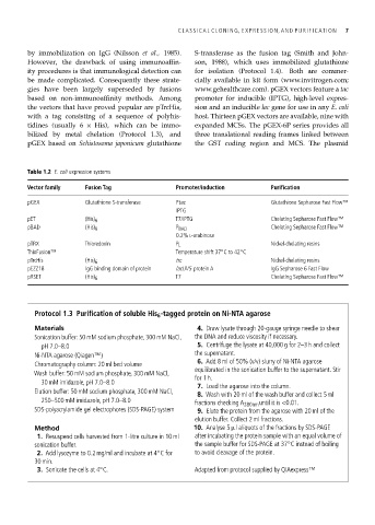
Table 1.2 E. coli expression systems
Vector family Fusion Tag Promoter/induction Purification
pGEX Glutathione S-transferase Ptac Glutathione Sepharose Fast Flow™
IPTG
pET (His) 6 T7/IPTG Chelating Sepharose Fast Flow™
pBAD (His) 6 P BAD Chelating Sepharose Fast Flow™
0.2% L-arabinose
pTRX Thioredoxin P L Nickel-chelating resins
◦
ThioFusion™ Temperature shift 37 Cto42 C
◦
pTrcHis (His) 6 trc Nickel-chelating resins
pEZZ18 IgG binding domain of protein lacUV5 protein A IgG Sepharose 6 Fast Flow
pRSET (His) 6 T7 Chelating Sepharose Fast Flow™
Protocol 1.3 Purification of soluble His 6 -tagged protein on Ni-NTA agarose
Materials 4. Draw lysate through 20-gauge syringe needle to shear
Sonication buffer: 50 mM sodium phosphate, 300 mM NaCl, the DNA and reduce viscosity if necessary.
pH 7.0–8.0 5. Centrifuge the lysate at 40,000 g for 2–3 h and collect
the supernatant.
Ni-NTA agarose (Qiagen™)
6. Add 8 ml of 50% (v/v) slurry of Ni-NTA agarose
Chromatography column: 20 ml bed volume
equilibrated in the sonication buffer to the supernatant. Stir
Wash buffer: 50 mM sodium phosphate, 300 mM NaCl,
for 1 h.
30 mM imidazole, pH 7.0–8.0
7. Load the agarose into the column.
Elution buffer: 50 mM sodium phosphate, 300 mM NaCl,
8. Wash with 20 ml of the wash buffer and collect 5 ml
250–500 mM imidazole, pH 7.0–8.0 fractions checking A 280nm until it is <0.01.
SDS-polyacrylamide gel electrophores (SDS-PAGE) system 9. Elute the protein from the agarose with 20 ml of the
elution buffer. Collect 2 ml fractions.
Method 10. Analyse 5 µl aliquots of the fractions by SDS-PAGE
1. Resuspend cells harvested from 1-litre culture in 10 ml after incubating the protein sample with an equal volume of
◦
sonication buffer. the sample buffer for SDS-PAGE at 37 C instead of boiling
2. Add lysozyme to 0.2 mg/ml and incubate at 4 C for to avoid cleavage of the protein.
◦
30 min.
◦
3. Sonicate the cells at 4 C. Adapted from protocol supplied by QIAexpress™