
Page 176 - Microtectonics
P. 176
6.2 · Veins 165
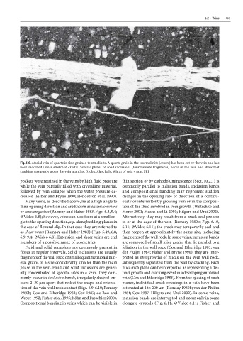
Fig. 6.6. Ataxial vein of quartz in fine-grained tourmalinite. A quartz grain in the tourmalinite (centre) has been cut by the vein and has
been modified into a stretched crystal. Several planes of solid inclusions (tourmalinite fragments) occur in the vein and show that
cracking was partly along the vein margins. Orobic Alps, Italy. Width of vein 4 mm. PPL
pockets were retained in the veins by high fluid pressure thin section or by cathodoluminescence (Sect. 10.2.1) is
while the vein partially filled with crystalline material, commonly parallel to inclusion bands. Inclusion bands
followed by vein collapse when the water pressure de- and compositional banding may represent sudden
creased (Fisher and Bryne 1990; Henderson et al. 1990). changes in the opening rate or direction of a continu-
Many veins, as described above, lie at a high angle to ously or intermittently growing vein or in the composi-
their opening direction and are known as extension veins tion of the fluid involved in vein growth (Wiltschko and
or tension gashes (Ramsay and Huber 1983; Figs. 6.8, 9.4; Morse 2001; Means and Li 2001; Hilgers and Urai 2002).
×Video 6.8); however, veins can also form at a small an- Alternatively, they may result from a crack-seal process
gle to the opening direction, e.g. along bedding planes in in or at the edge of the vein (Ramsay 1980b; Figs. 6.10,
the case of flexural slip. In that case they are referred to 6.11; ×Video 6.11); the crack may temporarily seal and
as shear veins (Ramsay and Huber 1983) (Figs. 5.49, 6.8, then reopen at approximately the same site, including
6.9, 9.4; ×Video 6.8). Extension and shear veins are end fragments of the wall rock. In some veins, inclusion bands
members of a possible range of geometries. are composed of small mica grains that lie parallel to a
Fluid and solid inclusions are commonly present in foliation in the wall rock (Cox and Etheridge 1983; van
fibres at regular intervals. Solid inclusions are usually der Pluijm 1984; Fisher and Bryne 1990); they are inter-
fragments of the wall rock, or small equidimensional min- preted as overgrowths of micas on the vein wall rock,
eral grains of a size considerably smaller than the main subsequently separated from the wall by cracking. Each
phase in the vein. Fluid and solid inclusions are gener- mica-rich plane can be interpreted as representing a dis-
ally concentrated at specific sites in a vein. They com- tinct growth and cracking event in a developing antitaxial
monly occur in inclusion bands, irregularly shaped sur- vein (Cox and Etheridge 1983). From the spacing of such
faces 2–30 µm apart that reflect the shape and orienta- planes, individual crack openings in a vein have been
tion of the vein-wall rock contact (Figs. 6.9, 6.10; Ramsay estimated at 4 to 200 µm (Ramsay 1980b; van der Pluijm
1980b; Cox and Etheridge 1983; Cox 1987; de Roo and 1984; Cox 1987; Hilgers and Urai 2002). In some veins,
Weber 1992; Fisher et al. 1995; Köhn and Passchier 2000). inclusion bands are interrupted and occur only in some
Compositional banding in veins which can be visible in elongate crystals (Fig. 6.11, ×Video 6.11; Fisher and