
Page 126 - Multidimensional Chromatography
P. 126
118 Multidimensional Chromatography
components from aqueous solutions are injected on to a reversed-phase column. A
similar outcome is achieved by adsorption chromatographic methods employing suit-
able solvent polarities. Subsequent elution with a stronger eluent will remove the
retained analyte on the first column and will start the separation procedure on the ana-
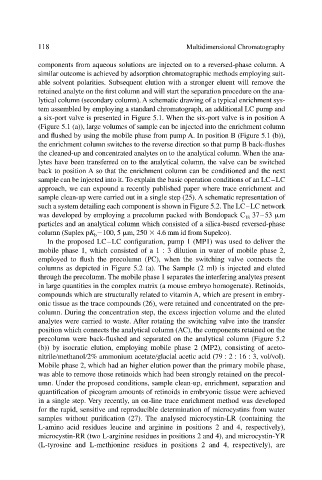
lytical column (secondary column). A schematic drawing of a typical enrichment sys-
tem assembled by employing a standard chromatograph, an additional LC pump and
a six-port valve is presented in Figure 5.1. When the six-port valve is in position A
(Figure 5.1 (a)), large volumes of sample can be injected into the enrichment column
and flushed by using the mobile phase from pump A. In position B (Figure 5.1 (b)),
the enrichment column switches to the reverse direction so that pump B back-flushes
the cleaned-up and concentrated analytes on to the analytical column. When the ana-
lytes have been transferred on to the analytical column, the valve can be switched
back to position A so that the enrichment column can be conditioned and the next
sample can be injected into it. To explain the basic operation conditions of an LC–LC
approach, we can expound a recently published paper where trace enrichment and
sample clean-up were carried out in a single step (25). A schematic representation of
such a system detailing each component is shown in Figure 5.2. The LC–LC network
was developed by employing a precolumn packed with Bondopack C 18 37–53 m
particles and an analytical column which consisted of a silica-based reversed-phase
column (Suplex pK b 100, 5 m, 250 4.6 mm id from Supelco).
In the proposed LC–LC configuration, pump 1 (MP1) was used to deliver the
mobile phase 1, which consisted of a 1 : 3 dilution in water of mobile phase 2,
employed to flush the precolumn (PC), when the switching valve connects the
columns as depicted in Figure 5.2 (a). The Sample (2 ml) is injected and eluted
through the precolumn. The mobile phase 1 separates the interfering analytes present
in large quantities in the complex matrix (a mouse embryo homogenate). Retinoids,
compounds which are structurally related to vitamin A, which are present in embry-
onic tissue as the trace compounds (26), were retained and concentrated on the pre-
column. During the concentration step, the excess injection volume and the eluted
analytes were carried to waste. After rotating the switching valve into the transfer
position which connects the analytical column (AC), the components retained on the
precolumn were back-flushed and separated on the analytical column (Figure 5.2
(b)) by isocratic elution, employing mobile phase 2 (MP2), consisting of aceto-
nitrile methanol 2% ammonium acetate glacial acetic acid (79 : 2 : 16 : 3, vol/vol).
Mobile phase 2, which had an higher elution power than the primary mobile phase,
was able to remove those retinoids which had been strongly retained on the precol-
umn. Under the proposed conditions, sample clean-up, enrichment, separation and
quantification of picogram amounts of retinoids in embryonic tissue were achieved
in a single step. Very recently, an on-line trace enrichment method was developed
for the rapid, sensitive and reproducible determination of microcystins from water
samples without purification (27). The analysed microcystin-LR (containing the
L-amino acid residues leucine and arginine in positions 2 and 4, respectively),
microcystin-RR (two L-arginine residues in positions 2 and 4), and microcystin-YR
(L-tyrosine and L-methionine residues in positions 2 and 4, respectively), are