
Page 149 - Optofluidics Fundamentals, Devices, and Applications
P. 149
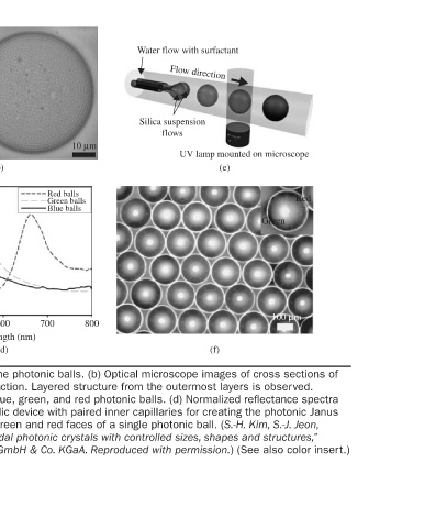
UV lamp mounted on microscope  (e)  Red  Green  100 μm


         Water flow with surfactant  Flow direction  Silica suspension  flows  (f)


                       10 μm                      800
                               Red balls  Green balls  Blue balls
                                                  700
                                                    Wavelength (nm)  (d) (a) Schematic illustration of optofluidic device for creating the photonic balls. (b) Optical microscope images of cross sections of
                                                  600
                           (b)                              emulsion droplets which contain 1-μm silica particles at 33% volume fraction. Layered structure from the outermost layers is observed.  (c) Optical microscope image of monodisperse emulsion droplets and blue, green, and red photonic balls. (d) Normalized r

                                                  500


                                                  400
                                             Reflectance (Arb. U.)

                                                  100 μm


          Water flow with surfactant  Flow direction  Silica suspension  flow  UV lamp mounted on microscope  (a)  Blue  Red  (c)


                                           Green
                                                          FIGURE 6-5


                                                                   125