
Page 274 -
P. 274
262 6 Structural Pattern Recoenition
Figure 6.13. An ECG signal, zoomed 4x, described by a piecewise approximation
using Chebychev norm with tolerance 20, with segments labelled using slope
thresholds of 5 and 22. Colour coding as in Figure 6.1 1.
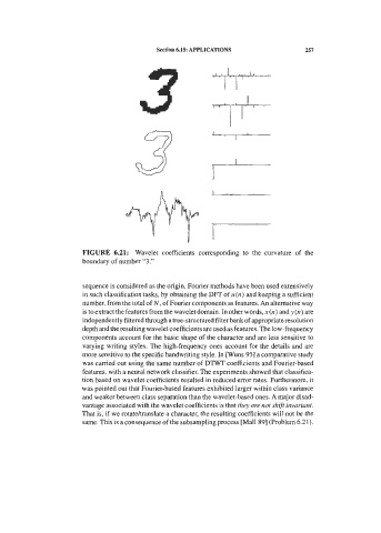
As an example, let us consider the distinction between negative P waves and Q
waves in an electrocardiographic signal, as shown in Figure 6.13. A brief
description of these waves can be found In section 1.3. P waves can be positive
and/or negative. Q waves, by definition, are negative. Figure 6.13 shows a negative
P wave, described by the string dU, followed by a Q wave, described by the same
string.
Figure 6.14. State-diagrams of tinite-state automata for the recognition of negative
' I
P waves (a) and Q waves (b).
The state-diagrams of finite-state slutonlata recognizing negative P waves and Q
waves are shown in Figure 6.14. Each arc in these diagrams is labelled with the
corresponding transition probability. Notice that rule (6-15) is satisfied. Notice also
that the state transition probabilities reflect the fact that a negative P wave is
usually less peaked than a Q wave.