
Page 127 - Visions of the Future Chemistry and Life Science
P. 127
116 M. C. H. VAN DER MEULEN AND P. J. PRENDERGAST
Epiphysis Metaphysis Diaphysis Metaphysis Epiphysis
Endosteum
Medullary Cortical Cancellous
Growth plate canal bone Periosteum bone
(a)
Osteoclast
Osteoblast
Lining
Cell
Osteocyte
Howship's
lacuna
Canaliculus
(b)
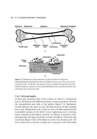
Figure 7.2. Schematics of bone anatomy: (a) the structure of a long bone
demonstrating the distribution of the two different tissue structures, cortical and
cancellous bone, and (b) the cells present in bone: osteoblasts, bone-forming cells
found on surfaces; osteocytes, bone cells embedded in the mineralised matrix; and
osteoclasts, bone-removing cells.
7.2.2 Cells and matrix
Cortical and cancellous bone tissue consists of cells in a mineralised
matrix. All skeletal cells differentiate from a common precursor cell pool:
the mesenchymal stem cells of the embryo (Figure 7.3). Mechanical
stimuli influence the mode of stem cell differentiation and the resulting
tissue type. Manipulation and control of stem cell differentiation holds
considerable promise in the field of tissue engineering and is receiving
much commercial and ethical attention. In addition to precursor cells,
three principal cell types are present in bone: osteoblasts, osteocytes, and
osteoclasts (Figure 7.2(b)). Osteoblasts are active bone-forming cells. All
bone surfaces are covered by a single layer of precursor cells and resting