
Page 108 - Algae Anatomy, Biochemistry, and Biotechnology
P. 108
Anatomy 91
these fibrils would act as channels for the extruded slime to flow along. Therefore, if the fibrils are
helically arranged, the cell will rotate as it glides; if the fibrils are aligned radially, the cell will
not rotate. In all species studied to date, this correlation is consistent, and provides a structural
explanation for why some species rotate as they glide while others do not.
In diatoms, motility is restricted to pennate species possessing a raphe. These diatoms display a
characteristic jerky movement forward or backward, with specie-specific path patterns. The general
velocity of their movement is 1–25 mm sec 21 , but they can accelerate up to 100–200 mm sec 21 .
Raphid diatoms possess an actin-based cytoskeletal system located just beneath the plasma
membrane at the raphe. Transmembrane components with an adhesive extracellular domain are
connected to these actin bundles, and their interaction is somehow involved in both adhesion
and motility mechanisms. Microtubules are also present in this region; in addition secretory ves-
icles containing polysaccharides often appear near the actin filaments at the raphe, providing the
mucilage strands that project from the raphe and adhere to the substratum during the gliding
process.
At least two models exist which provide reasonable explanation for diatom locomotion. In the
first model, a force applied to the transmembrane protein-actin connectors, parallel to the actin
bundles, would result in the movement of trasmembrane proteins through the cell and subsequent
movement of the cell in the opposite direction to the force. In the second model, the energy required
for motility would be generated by a conformational change of the adhesive mucilage on hydration
that occurs when it is secreted from the raphe. In this model, the actin bundles restrict the secretion
of mucilage to one end of the raphe, which generates a net force moving the cell over the site of
secretion. In both models, the secreted mucilage plays a central role either by providing traction
to translate the force into cell movement or by generating the energy through conformational
changes on hydration.
A slow gliding movement over solid substrata has been observed in Porphyridium sp.
(Rhodophyta) and in some desmids (Chlorophyta). In Porphyridium, the mucilage produced in
mucilage sacs located inside the cell is excreted through the membrane. In desmids mucilage is
excreted through the cell wall by flask-shaped pores. As they move, these gliding cells leave
behind a fibrillar mucilaginous trail, whose swelling by water pushes the cells forward.
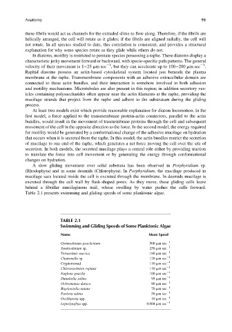
Table 2.1 presents swimming and gliding speeds of some planktonic algae.
TABLE 2.1
Swimming and Gliding Speeds of Some Planktonic Algae
Name Mean Speed
Gymnodinium gracilentum 500 mm sec 21
Symbiodinium sp. 250 mm sec 21
Tetraselmis suecica 180 mm sec 21
Chattonella sp. 120 mm sec 21
Cryptomonad 110 mm sec 21
Chlorarachnion reptans 110 mm sec 21
Euglena gracilis 100 mm sec 21
Dunaliella salina 95 mm sec 21
Ochromonas danica 80 mm sec 21
Bigelowiella natans 70 mm sec 21
Pavlova salina 50 mm sec 21
Oscillatoria spp. 10 mm sec 21
Leptolyngbya spp. 0.004 mm sec 21