
Page 113 - Algae Anatomy, Biochemistry, and Biotechnology
P. 113
96 Algae: Anatomy, Biochemistry, and Biotechnology
On the assumption that photoreception is inseparable from the presence of photoreceptive proteins
but not necessarily from the presence of an eyespot, we will group photoreceptive systems into
three main types. The description will include also those algae with well documented eyespot
but only presumptive photoreceptor localization.
TYPE I
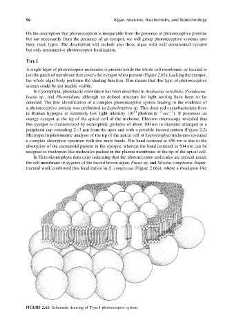
A single layer of photoreceptor molecules is present inside the whole cell membrane, or located in
just the patch of membrane that covers the eyespot when present (Figure 2.65). Lacking the eyespot,
the whole algal body performs the shading function. This means that this type of photoreceptive
system could be not readily visible.
In Cyanophyta, phototactic orientation has been described in Anabaena variabilis, Pseudoana-
baena sp., and Phormidium, although no defined structure for light sensing have been so far
detected. The first identification of a complex photoreceptive system leading to the evidence of
a photoreceptive protein was performed in Leptolyngbya sp. This deep red cyanobacterium lives
13 22 21
in Roman hypogea at extremely low light intensity (10 photons m sec ). It possesses an
orange eyespot at the tip of the apical cell of the trichome. Electron microscopy revealed that
this eyespot is characterized by osmiophilic globules of about 100 nm in diameter arranged in a
peripheral cap extending 2–3 mm from the apex and with a possible layered pattern (Figure 2.2)
Microspectrophotometric analysis of the tip of the apical cell of Leptolyngbya trichomes revealed
a complex absorption spectrum with two main bands. The band centered at 456 nm is due to the
absorption of the carotenoid present in the eyespot, whereas the band centered at 504 nm can be
assigned to rhodopsin-like molecules packed in the plasma membrane of the tip of the apical cell.
In Heterokontophyta data exist indicating that the photoreceptor molecules are present inside
the cell membrane of zygotes of the fucoid brown algae, Fucus sp. and Silvetia compressa. Exper-
imental work confirmed this localization in S. compressa (Figure 2.66a), where a rhodopsin-like
FIGURE 2.65 Schematic drawing of Type I photoreceptor system.