
Page 114 - Algae Anatomy, Biochemistry, and Biotechnology
P. 114
Anatomy 97
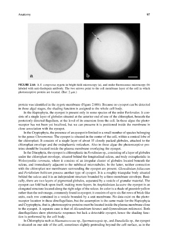
FIGURE 2.66 A S. compressa zygote in bright field microscopy (a), and under fluorescence microscopy (b)
labeled with anti-rhodopsin antibody. The two arrows point to the cell membrane layer of the cell in which
photoreceptive protein are located. (Bar: 2 mm.)
protein was identified in the zygote membrane (Figure 2.66b). Because no eyespot can be detected
in these algal stages, the shading function is assigned to the whole cell body.
In the Haptophyta, the eyespot is present only in some species of the order Pavlovales. It con-
sists of a single layer of globules situated at the anterior end of one of the chloroplast, beneath the
posteriorly directed flagellum, at the level of its emersion from the cell. In these algae the photo-
receptor has not been yet localized, but we can presume it is positioned inside the membrane in
close association with the eyespot.
In the Cryptophyta, the presence of an eyespot is limited to a small number of species belonging
to the genus Chroomonas. The eyespot is situated in the center of the cell, within a conical lobe of
the chloroplast. It consists of a single layer of about 35 closely packed globules, attached to the
chloroplast envelope and the endoplasmic reticulum. Also in these algae the photoreceptive pro-
teins should be located inside the plasma membrane overlaying the eyespot.
In the Dinophyta, the eyespot is chloroplastic in Peridiniun sp., consisting of a layer of globules
under the chloroplast envelope, situated behind the longitudinal sulcus, and truly extraplastidic in
Woloszynskia coronata, where it consists of an irregular cluster of globules located beneath the
sulcus, and immediately adjacent to the subthecal microtubules. In the latter, neither connection
with the chloroplast nor membranes surrounding the eyespot are present. Glenodinium foliaceum
and Peridinium balticum possess another type of eyespot. It is a roughly triangular body situated
behind the sulcus and it is an independent structure bounded by a three-membrane envelope. Basi-
cally, there are two layers of pigmented globules, separated by a vesicle of granular material. The
eyespot can fold back upon itself, making more layers. In Amphidinium lacustre the eyespot is an
elongated structure located along the right edge of the sulcus. Its color is a shade of greenish-yellow
rather than the red-orange, commonly found in eyespot; it consists of up to six flat rows of brick-like
units, each row contained in a vesicle bounded by a unit membrane. No data exist on the photo-
receptor location in these dinoflagellates, but the assumption is the same made for the Haptophyta
and Cryptophyta, that is, photoreceptive proteins must be located inside the plasma membrane close
to the eyespot. A separate case is that of Alexandrium hiranoi and Gymnodinium mikimotoi. Both
dinoflagellates show phototactic responses but lack a detectable eyespot, hence the shading func-
tion is performed by the cell body.
In Chlorophyta such as Haematococcus sp., Spermatozopsis sp., and Dunaliella sp., the eyespot
is situated on one side of the cell, sometimes slightly protruding beyond the cell surface, as in the