
Page 117 - Algae Anatomy, Biochemistry, and Biotechnology
P. 117
100 Algae: Anatomy, Biochemistry, and Biotechnology
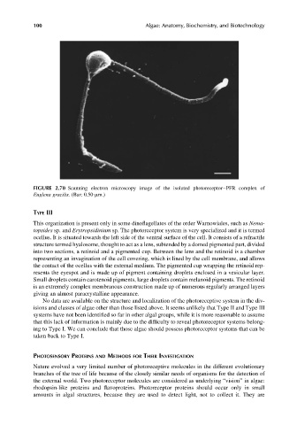
FIGURE 2.70 Scanning electron microscopy image of the isolated photoreceptor–PFR complex of
Euglena gracilis. (Bar: 0.50 mm.)
TYPE III
This organization is present only in some dinoflagellates of the order Warnowiales, such as Nema-
topsides sp. and Erytropsidinium sp. The photoreceptor system is very specialized and it is termed
ocellus. It is situated towards the left side of the ventral surface of the cell. It consists of a refractile
structure termed hyalosome, thought to act as a lens, subtended by a domed pigmented part, divided
into two sections, a retinoid and a pigmented cup. Between the lens and the retinoid is a chamber
representing an invagination of the cell covering, which is lined by the cell membrane, and allows
the contact of the ocellus with the external medium. The pigmented cup wrapping the retinoid rep-
resents the eyespot and is made up of pigment containing droplets enclosed in a vesicular layer.
Small droplets contain carotenoid pigments, large droplets contain melanoid pigments. The retinoid
is an extremely complex membranous construction made up of numerous regularly arranged layers
giving an almost paracrystalline appearance.
No data are available on the structure and localization of the photoreceptive system in the div-
isions and classes of algae other than those listed above. It seems unlikely that Type II and Type III
systems have not been identified so far in other algal groups, while it is more reasonable to assume
that this lack of information is mainly due to the difficulty to reveal photoreceptor systems belong-
ing to Type I. We can conclude that those algae should possess photoreceptor systems that can be
taken back to Type I.
PHOTOSENSORY PROTEINS AND METHODS FOR THEIR INVESTIGATION
Nature evolved a very limited number of photoreceptive molecules in the different evolutionary
branches of the tree of life because of the closely similar needs of organisms for the detection of
the external world. Two photoreceptor molecules are considered as underlying “vision” in algae:
rhodopsin-like proteins and flavoproteins. Photoreceptor proteins should occur only in small
amounts in algal structures, because they are used to detect light, not to collect it. They are