
Page 126 - Algae Anatomy, Biochemistry, and Biotechnology
P. 126
Anatomy 109
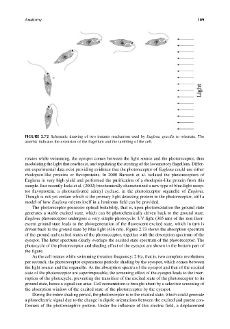
FIGURE 2.72 Schematic drawing of two instants mechanism used by Euglena gracilis to orientate. The
asterisk indicates the extension of the flagellum and the tumbling of the cell.
rotates while swimming, the eyespot comes between the light source and the photoreceptor, thus
modulating the light that reaches it, and regulating the steering of the locomotory flagellum. Differ-
ent experimental data exist providing evidence that the photoreceptor of Euglena could use either
rhodopsin-like proteins or flavoproteins. In 2000 Barsanti et al. isolated the photoreceptors of
Euglena in very high yield and performed the purification of a rhodopsin-like protein from this
sample. Just recently Iseki et al. (2002) biochemically characterized a new type of blue-light recep-
tor flavoprotein, a photoactivated adenyl cyclase, in the photoreceptor organelle of Euglena.
Though is not yet certain which is the primary light detecting protein in the photoreceptor, still a
model of how Euglena orients itself in a luminous field can be provided.
The photoreceptor possesses optical bistability, that is, upon photoexcitation the ground state
generates a stable excited state, which can be photochemically driven back to the ground state.
Euglena photoreceptor undergoes a very simple photocycle: UV light (365 nm) of the non-fluor-
escent ground state leads to the photogeneration of the fluorescent excited state, which in turn is
driven back to the ground state by blue light (436 nm). Figure 2.73 shows the absorption spectrum
of the ground and excited states of the photoreceptor, together with the absorption spectrum of the
eyespot. The latter spectrum clearly overlaps the excited state spectrum of the photoreceptor. The
photocycle of the photoreceptor and shading effect of the eyespot are shown in the bottom part of
the figure.
As the cell rotates while swimming (rotation frequency: 2 Hz, that is, two complete revolutions
per second), the photoreceptor experiences periodic shading by the eyespot, which comes between
the light source and the organelle. As the absorption spectra of the eyespot and that of the excited
state of the photoreceptor are superimposable, the screening effect of the eyespot leads to the inter-
ruption of the photocycle, preventing the transition of the excited state of the photoreceptor to its
ground state, hence a signal can arise. Cell reorientation is brought about by a selective screening of
the absorption window of the excited state of the photoreceptor by the eyespot.
During the entire shading period, the photoreceptor is in the excited state, which could generate
a photoelectric signal due to the change in dipole orientations between the excited and parent con-
formers of the photoreceptive protein. Under the influence of this electric field, a displacement