
Page 265 - Algae Anatomy, Biochemistry, and Biotechnology
P. 265
248 Algae: Anatomy, Biochemistry, and Biotechnology
chamber is 0.1 mm deep; hence each grid holds exactly 64 10 23 ml of sample. You have the choice
of counting the algae in the entire grid; counting algae in only one of the 16 squares, then multiplying by
16; or counting algae in an even smaller area and multiplying accordingly.
When counting cell in the entire grid, that is, 16 fields, the cell concentration is calculated
according to the following formula:
C 10 3
1
Number of cells mL ¼ (6:4)
64
where C is the number of cells counted.
Counts of about 30 cells per field are desirable for accuracy. If there are more than 30 cells
per field, dilute the sample, or count algae in a lower number of fields and multiply. As for the
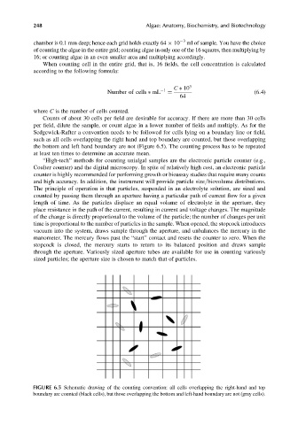
Sedgewick-Rafter a convention needs to be followed for cells lying on a boundary line or field,
such as all cells overlapping the right hand and top boundary are counted, but those overlapping
the bottom and left hand boundary are not (Figure 6.5). The counting process has to be repeated
at least ten times to determine an accurate mean.
“High-tech” methods for counting unialgal samples are the electronic particle counter (e.g.,
Coulter counter) and the digital microscopy. In spite of relatively high cost, an electronic particle
counter is highly recommended for performing growth or bioassay studies that require many counts
and high accuracy. In addition, the instrument will provide particle size/biovolume distributions.
The principle of operation is that particles, suspended in an electrolyte solution, are sized and
counted by passing them through an aperture having a particular path of current flow for a given
length of time. As the particles displace an equal volume of electrolyte in the aperture, they
place resistance in the path of the current, resulting in current and voltage changes. The magnitude
of the change is directly proportional to the volume of the particle; the number of changes per unit
time is proportional to the number of particles in the sample. When opened, the stopcock introduces
vacuum into the system, draws sample through the aperture, and unbalances the mercury in the
manometer. The mercury flows past the “start” contact and resets the counter to zero. When the
stopcock is closed, the mercury starts to return to its balanced position and draws sample
through the aperture. Variously sized aperture tubes are available for use in counting variously
sized particles; the aperture size is chosen to match that of particles.
FIGURE 6.5 Schematic drawing of the counting convention: all cells overlapping the right-hand and top
boundary are counted (black cells), but those overlapping the bottom and left-hand boundary are not (gray cells).