
Page 58 - Algae Anatomy, Biochemistry, and Biotechnology
P. 58
Anatomy 41
underlain by base-plate organic scales formed inside Golgi vesicles. However, holococcolith calcifi-
cation is an extracellular process. Experimental evidences revealed that calcification occurs in a single
highly regulated space outside the cell membrane, but directly above the stack of Golgi vesicles. This
extracellular compartment is covered by a delicate organic envelope or “skin.” The cell secretes
calcite that fills the space between the skin and the base-plate scales. The coccosphere grows pro-
gressively outward from this position. As a consequence of the different biomineralization strategies,
heterococcoliths are more robust than the smaller and more delicate holococcoliths.
Coccolithophorids, together with corals and foraminifera, are responsible for the bulk of
oceanic calcification. Their role in the formation of marine sediment and the impact their
blooms may exert on climate change will be discussed in Chapter 4.
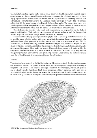
Members of the Chrysophyceae (Heterokontophyta) such as Synura sp. and Mallomonas sp. are
covered by armor of silica scales, with a very complicated structure. Synura scales consists of a
perforated basal plate provided with ribs, spines, and other ornamentation (Figure 2.8). In
Mallomonas, scales may bear long, complicated bristles (Figure 2.9). Several scale types are pro-
duced in the same cell and deposited on the surface in a definite sequence, following an imbricate,
often screw-like pattern. Silica scales are produced internally in deposition vesicles formed by the
chrysoplast endoplasmic reticulum, which function as moulds for the scales. Golgi body vesicles
transporting material fuse with the scale-producing vesicles. Once formed the scale is extruded
from the cell and brought into correct position on the cell surface.
Frustule
This structure is present only in the Bacillariophyceae (Heterokontophyta). The frustule is an ornate
cell membrane made of amorphous hydrated silica, which displays intricate patterns and designs
unique to each species. This silicified envelope consists of two overlapping valves, an epitheca
and a slightly smaller hypotheca. Each theca comprises a highly patterned valve and one or
more girdle bands (cingula) that extend around the circumference of the cell, forming the region
of theca overlay. Extracellular organic coats envelop the plasma membrane under the siliceous
FIGURE 2.8 Ornamented body scale of Synura petersenii.