
Page 61 - Algae Anatomy, Biochemistry, and Biotechnology
P. 61
44 Algae: Anatomy, Biochemistry, and Biotechnology
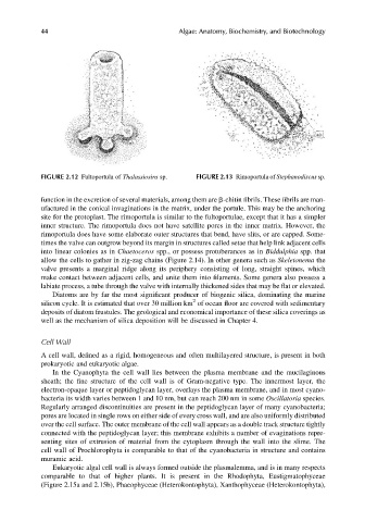
FIGURE 2.12 Fultoportula of Thalassiosira sp. FIGURE 2.13 Rimoportula of Stephanodiscus sp.
function in the excretion of several materials, among them are b-chitin fibrils. These fibrils are man-
ufactured in the conical invaginations in the matrix, under the portule. This may be the anchoring
site for the protoplast. The rimoportula is similar to the fultoportulae, except that it has a simpler
inner structure. The rimoportula does not have satellite pores in the inner matrix. However, the
rimoportula does have some elaborate outer structures that bend, have slits, or are capped. Some-
times the valve can outgrow beyond its margin in structures called setae that help link adjacent cells
into linear colonies as in Chaetoceros spp., or possess protuberances as in Biddulphia spp. that
allow the cells to gather in zig-zag chains (Figure 2.14). In other genera such as Skeletonema the
valve presents a marginal ridge along its periphery consisting of long, straight spines, which
make contact between adjacent cells, and unite them into filaments. Some genera also possess a
labiate process, a tube through the valve with internally thickened sides that may be flat or elevated.
Diatoms are by far the most significant producer of biogenic silica, dominating the marine
2
silicon cycle. It is estimated that over 30 million km of ocean floor are covered with sedimentary
deposits of diatom frustules. The geological and economical importance of these silica coverings as
well as the mechanism of silica deposition will be discussed in Chapter 4.
Cell Wall
A cell wall, defined as a rigid, homogeneous and often multilayered structure, is present in both
prokaryotic and eukaryotic algae.
In the Cyanophyta the cell wall lies between the plasma membrane and the mucilaginous
sheath; the fine structure of the cell wall is of Gram-negative type. The innermost layer, the
electron-opaque layer or peptidoglycan layer, overlays the plasma membrane, and in most cyano-
bacteria its width varies between 1 and 10 nm, but can reach 200 nm in some Oscillatoria species.
Regularly arranged discontinuities are present in the peptidoglycan layer of many cyanobacteria;
pores are located in single rows on either side of every cross wall, and are also uniformly distributed
over the cell surface. The outer membrane of the cell wall appears as a double track structure tightly
connected with the peptidoglycan layer; this membrane exhibits a number of evaginations repre-
senting sites of extrusion of material from the cytoplasm through the wall into the slime. The
cell wall of Prochlorophyta is comparable to that of the cyanobacteria in structure and contains
muramic acid.
Eukaryotic algal cell wall is always formed outside the plasmalemma, and is in many respects
comparable to that of higher plants. It is present in the Rhodophyta, Eustigmatophyceae
(Figure 2.15a and 2.15b), Phaeophyceae (Heterokontophyta), Xanthophyceae (Heterokontophyta),