
Page 65 - Algae Anatomy, Biochemistry, and Biotechnology
P. 65
48 Algae: Anatomy, Biochemistry, and Biotechnology
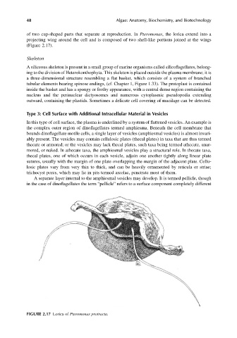
of two cup-shaped parts that separate at reproduction. In Pteromonas, the lorica extend into a
projecting wing around the cell and is composed of two shell-like portions joined at the wings
(Figure 2.17).
Skeleton
A siliceous skeleton is present in a small group of marine organisms called silicoflagellates, belong-
ing to the division of Heterokonthophyta. This skeleton is placed outside the plasma membrane; it is
a three-dimensional structure resembling a flat basket, which consists of a system of branched
tubular elements bearing spinose endings, (cf. Chapter 1, Figure 1.33). The protoplast is contained
inside the basket and has a spongy or frothy appearance, with a central dense region containing the
nucleus and the perinuclear dictyosomes and numerous cytoplasmic pseudopodia extending
outward, containing the plastids. Sometimes a delicate cell covering of mucilage can be detected.
Type 3: Cell Surface with Additional Intracellular Material in Vesicles
In this type of cell surface, the plasma is underlined by a system of flattened vesicles. An example is
the complex outer region of dinoflagellates termed amphiesma. Beneath the cell membrane that
bounds dinoflagellate motile cells, a single layer of vesicles (amphiesmal vesicles) is almost invari-
ably present. The vesicles may contain cellulosic plates (thecal plates) in taxa that are thus termed
thecate or armored; or the vesicles may lack thecal plates, such taxa being termed athecate, unar-
mored, or naked. In athecate taxa, the amphiesmal vesicles play a structural role. In thecate taxa,
thecal plates, one of which occurs in each vesicle, adjoin one another tightly along linear plate
sutures, usually with the margin of one plate overlapping the margin of the adjacent plate. Cellu-
losic plates vary from very thin to thick, and can be heavily ornamented by reticula or striae;
trichocyst pores, which may lie in pits termed areolae, penetrate most of them.
A separate layer internal to the amphiesmal vesicles may develop. It is termed pellicle, though
in the case of dinoflagellates the term “pellicle” refers to a surface component completely different
FIGURE 2.17 Lorica of Pteromonas protracta.