
Page 70 - Algae Anatomy, Biochemistry, and Biotechnology
P. 70
Anatomy 53
FIGURE 2.21 Transmission electron microscopy image of the surface of Euglena gracilis in transverse
section, showing the three different structural levels of the pellicle. Arrows point to the first level (mucus
coating); a square bracket localizes the second level (ridges and grooves); arrowheads point the third level
(microtubules). (Bar: 0.10 mm.)
First Level
A dense irregular layer of mucilaginous glycoprotein covers the external surface of the cell. It has a
fuzzy texture that, however, has a somehow ordered structure of orientated threads. Mucilage
bodies present beneath the cell surface secret the mucilaginous glycoproteins. The consolidation
of the secretory products and their arrangement at one pole or around the periphery of the cell
leads to the formation of peduncles (stalks of fixation) and other enveloping structures homologous
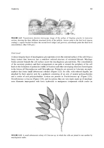
to the loricas of Chrysophyceae and Chlorophyceae. Peduncles are present in Colacium, an eugle-
nophyte that forms small arborescent colonies (Figure 2.22). Its cells, with reduced flagella, are
attached by their anterior pole by a peduncle consisting of an axis of neutral polysaccharides
and a cortex of acid polysaccharides. Loricas are present in Trachelomonas sp. (Figure 2.23),
Strombomonas verrucosa (Figure 2.24), and Ascoglena; they are very rigid, made up of mucilagi-
nous filaments impregnated with ferric hydroxide or manganese compounds which confer an
FIGURE 2.22 A small arborescent colony of Colacium sp. in which the cells are joined to one another by
mucilaginous stalks.