
Page 71 - Algae Anatomy, Biochemistry, and Biotechnology
P. 71
54 Algae: Anatomy, Biochemistry, and Biotechnology
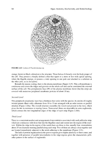
FIGURE 2.23 Lorica of Trachelomonas sp.
orange, brown to black coloration to the structure. These loricas fit loosely over the body proper of
the cell. They possess a sharply defined collar that tapers to a more or less wide apical opening,
where the flagella emerge, or possess a wide opening in one pole and attached to a substrate at
the other pole, as in Ascoglena.
Beneath the mucus coating, there is the plasma membrane (Figure 2.25). This cell membrane is
continuous and covers the ridges and grooves on the whole cell and can be considered the external
surface of the cell. The protoplasmic face (PF) of the plasma membrane shows that the strips are
covered with numerous peripheral membrane proteins of about 10 nm.
Second Level
This peripheral cytoplasmic layer has a thickness that varies with the species. It consists of roughly
twisted proteic fibers with a diameter from 10 to 15 nm arranged with an order texture or parallel
striation (Figure 2.26a). The overall structure resembles the wired soul present in the tires, which
gives the tire its resistance to tearing forces. Transversal fibers are detectable in some euglenoids,
which connect the two longitudinal edges of the ridge of each strip (Figure 2.26b).
Third Level
There is a consistent number and arrangement of microtubules associated with each pellicular strip,
which are continuous with those that line the flagellar canal and extend into the region of the reser-
voir. Within the ridge in the region of the notch there are three to five, usually four, microtubules
about 25 nm diameter running parallel along each strip. Two of these are always close together and
are located immediately adjacent to the notch adhering to the membrane (Figure 2.21).
The lack of protein organization in the groove regions gives higher plasticity to these zones, and
together with presence of parallel microtubules in the ridge regions gives the characteristic pelli-
cular pattern to the surface of euglenoids.