
Page 73 - Algae Anatomy, Biochemistry, and Biotechnology
P. 73
56 Algae: Anatomy, Biochemistry, and Biotechnology
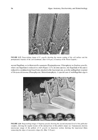
FIGURE 2.25 Deep-etching image of E. gracilis showing the mucus coating of the cell surface and the
protoplasmic fracture of the cell membrane. (Bar: 0.10 mm.) (Courtesy of Dr. Pietro Lupetti.)
second flagellum, as in Mantoniella squamata (Prasinophyceae, Chlorophyta) or Euglena gracilis,
where one flagellum is reduced to a stub (Figure 2.27); in some species, one flagellum of the pair is
reduced to a nonfunctional basal body attached to the functional one, as in the uniflagellate swarmer
of Dictyota dichotoma (Phaeophyceae, Heterokontophyta). A special case of multiflagellate alga is
FIGURE 2.26 Deep-etching image of Euglena gracilis showing the second structural level of the pellicular
complex, showing the regular texture of the internal face of the pellicle stripes (a). Transmission electron
microscopy image of the pellicle of E. gracilis in transverse section showing the transversal fibers
connecting the edges of successive ridges (b). (Bar: 0.10 mm.)