
Page 77 - Algae Anatomy, Biochemistry, and Biotechnology
P. 77
60 Algae: Anatomy, Biochemistry, and Biotechnology
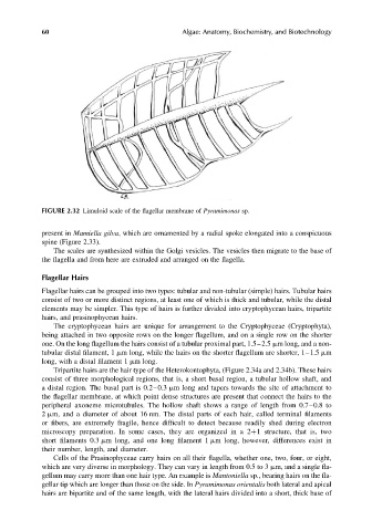
FIGURE 2.32 Limuloid scale of the flagellar membrane of Pyramimonas sp.
present in Mamiella gilva, which are ornamented by a radial spoke elongated into a conspicuous
spine (Figure 2.33).
The scales are synthesized within the Golgi vesicles. The vesicles then migrate to the base of
the flagella and from here are extruded and arranged on the flagella.
Flagellar Hairs
Flagellar hairs can be grouped into two types: tubular and non-tubular (simple) hairs. Tubular hairs
consist of two or more distinct regions, at least one of which is thick and tubular, while the distal
elements may be simpler. This type of hairs is further divided into cryptophycean hairs, tripartite
hairs, and prasinophycean hairs.
The cryptophycean hairs are unique for arrangement to the Cryptophyceae (Cryptophyta),
being attached in two opposite rows on the longer flagellum, and on a single row on the shorter
one. On the long flagellum the hairs consist of a tubular proximal part, 1.5–2.5 mm long, and a non-
tubular distal filament, 1 mm long, while the hairs on the shorter flagellum are shorter, 1–1.5 mm
long, with a distal filament 1 mm long.
Tripartite hairs are the hair type of the Heterokontophyta, (Figure 2.34a and 2.34b). These hairs
consist of three morphological regions, that is, a short basal region, a tubular hollow shaft, and
a distal region. The basal part is 0.2–0.3 mm long and tapers towards the site of attachment to
the flagellar membrane, at which point dense structures are present that connect the hairs to the
peripheral axoneme microtubules. The hollow shaft shows a range of length from 0.7–0.8 to
2 mm, and a diameter of about 16 nm. The distal parts of each hair, called terminal filaments
or fibers, are extremely fragile, hence difficult to detect because readily shed during electron
microscopy preparation. In some cases, they are organized in a 2þ1 structure, that is, two
short filaments 0.3 mm long, and one long filament 1 mm long, however, differences exist in
their number, length, and diameter.
Cells of the Prasinophyceae carry hairs on all their flagella, whether one, two, four, or eight,
which are very diverse in morphology. They can vary in length from 0.5 to 3 mm, and a single fla-
gellum may carry more than one hair type. An example is Mantoniella sp., bearing hairs on the fla-
gellar tip which are longer than those on the side. In Pyramimonas orientalis both lateral and apical
hairs are bipartite and of the same length, with the lateral hairs divided into a short, thick base of