
Page 124 - Advances in Biomechanics and Tissue Regeneration
P. 124
120 7. MULTISCALE NUMERICAL SIMULATION OF HEART ELECTROPHYSIOLOGY
et al. [40] for human atria, ten Tusscher et al. [8, 41], O’Hara et al. [42], and Carro et al. [43] for human ventricles, Luo
and Rudy [44, 45] for guinea pig ventricles, and Shannon et al. [46] for rabbit ventricles, among others.
In this work, we have implemented a number of AP models, namely: Maleckar et al. (MA09) and Nygren et al.
(NY98) for atria, ten Tusscher et al. (TP06) and O’Hara et al (OH11) for human ventricles, Stewart et al. (ST99) for
Purkinje cells, and Bueno-Orovio et al. (BV08) as a type of phenomenological model. The TP06 model will be exten-
sively used in Chapter 4 to study the acute ischemic heart. In the following, the basic structure of a modern AP model is
described in detail.
7.2.3.1 Structure of an Action Potential Model
As known, it is possible to reproduce the characteristics of AP with simple models. However, an important objective
in the modelization of physiological phenomena is to research how the changes in the cell physiology affect the tissue
and finally, the studied organ. For this purpose, it is necessary that the models study the cell physiology from the mem-
brane to the ionic channels, which set up the gates for the exchange between the intracellular and extracellular media,
including the dynamic mechanism in the cytoplasm.
7.2.3.2 The Cell Membrane
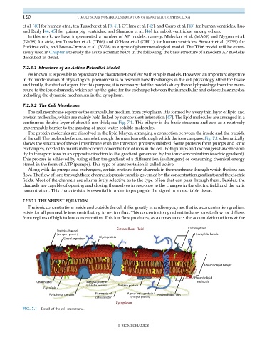
The cell membrane separates the extracellular medium from cytoplasm. It is formed by a very thin layer of lipid and
protein molecules, which are mainly held linked by noncovalent interaction [47]. The lipid molecules are arranged in a
continuous double layer of about 5 nm thick; see Fig. 7.1. This bilayer is the basic structure and acts as a relatively
impermeable barrier to the passing of most water-soluble molecules.
The protein molecules are dissolved in the lipid bilayer, arranging a connection between the inside and the outside
of the cell. The molecules form channels through the membrane through which the ions can pass. Fig. 7.1 schematically
shows the structure of the cell membrane with the transport proteins imbibed. Some proteins form pumps and ionic
exchangers, needed to maintain the correct concentration of ions in the cell. Both pumps and exchangers have the abil-
ity to transport ions in an opposite direction to the gradient generated by the ionic concentration (electric gradient).
This process is achieved by using either the gradient of a different ion (exchangers) or consuming chemical energy
stored in the form of ATP (pumps). This type of transportation is called active.
Along with the pumps and exchangers, certain proteins form channels in the membrane through which the ions can
flow. The flow of ions through these channels is passive and is governed by the concentration gradients and the electric
fields. Most of the channels are alternatively selective as to the type of ion that can pass through them. Besides, the
channels are capable of opening and closing themselves in response to the changes in the electric field and the ionic
concentration. This characteristic is essential in order to propagate the signal in an excitable tissue.
7.2.3.2.1 THE NERNST EQUATION
The ionic concentrations inside and outside the cell differ greatly in cardiomyocytes, that is, a concentration gradient
exists for all permeable ions contributing to net ion flux. This concentration gradient induces ions to flow, or diffuse,
from regions of high to low concentration. This ion flow produces, as a consequence, the accumulation of ions at the
FIG. 7.1 Detail of the cell membrane.
I. BIOMECHANICS