
Page 194 - Advances in Textile Biotechnology
P. 194
Enzymatic treatment of wool and silk fi bres 175
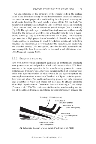
An understanding of the structure of the cuticlar cells in the surface
scales of the fibres is necessary for the development of successful enzymatic
processes for wool preparation and finishing including wool scouring and
shrink-resist finishing. The wool cuticle is about 400 to 500 nm thick. The
cuticlar cells comprise an endocuticle (120 to 180 nm thick), an exocuticle
(150 to 200 nm thick) and an outermost epicuticle layer (5 to 7 nm thick)
(Fig. 8.2). The epicuticle layer contains 18-methyleicosanoic acid covalently
bonded to the surface of wool fibre via a thioester bond to form a hydro-
phobic barrier (a fatty acid monolayer called the F-layer). The exocuticle
layer contains a high proportion of crosslinked disulfide and isopeptide
bonds, resulting in resistance to attack by alkaline agents and proteolytic
enzymes. The endocuticle, a layer lying below the exocuticle, has a relatively
low crosslink density (3% half-cystine) and thus is easily permeable and
more susceptible than the exocuticle to chemical attack (Feldtman et al.,
1983; Mori and Inagaki, 2006).
8.3.2 Enzymatic scouring
Raw wool fibres contain significant quantities of contaminants including
wool greases, suint and soil particles which could be up to about 60%. Wool
scouring is the major operation in the manufacturing process to remove
contaminants from raw wool. There are several methods of scouring wool
either with aqueous solution or with solvents. In the aqueous system, the
scouring line consists of a number of bowls of hot liquor containing water,
detergent and alkali. The traditional scouring process not only consumes
large quantities of water and energy but also leads to effl uent discharge
−1
with a high organic content, COD from 5000 to 150 000 mg L , and sludge
(Pearson et al., 1976). The environmental impact of wool-scouring and the
cost of the effluent treatment and sludge disposal increasingly concern the
Epicuticle (12% half-cystine)
Exocuticle ‘A’
(35% half-cystine)
Exocuticle ‘B’
(15% half-cystine)
Endocuticle
(3% half-cystine)
Intercellular cement
(1% half-cystine)
8.2 Schematic diagram of wool cuticle (Feldtman et al., 1983).
© Woodhead Publishing Limited, 2010