
Page 54 - Algae
P. 54
Anatomy 37
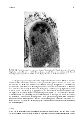
FIGURE 2.2 Transmission electron microscopy image of the apical cell of Leptolyngbya spp. trichome in
longitudinal section. The arrows point to the mucilaginous sheath of this cyanobacterium. Inside the cell
osmiophylic eyespot globules are present. (Bar: 0.15 mm). (Courtesy of Dr. Patrizia Albertano.)
In eukaryotic algae, mucilages and sheaths are present in diverse divisions. The most common
occurence of this extracellular material is in the algae palmelloid phases, in which non-motile cells
are embedded in a thick, more or less stratified sheath of mucilage. This phase is so-called because
it occurs in the genus Palmella (Chlorophyceae), but it occurs also in other members of the same
class, such as Asterococcus sp., Hormotila sp., Spirogyra sp., and Gleocystis sp. A palmelloid phase
is present also in Chroomonas sp. (Cryptophyceae) and in Gleodinium montanum vegetative cells
(Dynophyceae) and in Euglena gracilis (Euglenophyceae) (Figure 2.3). Less common are the cases
in which filaments are covered by continuous tubular layers of mucilages and sheath. It occurs in
the filaments of Geminella sp. (Chlorophyceae). A more specific covering exists in the filaments of
Phaeothamnion sp. (Chrysophyceae), because under certain growth conditions, cells of the fila-
ments dissociate and produce a thick mucilage that surround them in a sort of colony resembling
the palmelloid phase.
Scales
Scales can be defined as organic or inorganic surface structures of distinct size and shape. Scales
can be distributed individually or arranged in a pattern sometimes forming an envelope around