
Page 40 - Anthropometry, Apparel Sizing and Design
P. 40
36 Anthropometry, Apparel Sizing and Design
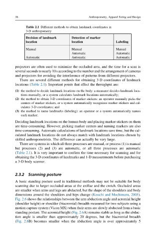
Table 2.1 Different methods to obtain landmark coordinates in
3-D anthropometry
Decision of landmark Detection of marker
location location Labeling
Manual Manual Manual
Automatic Automatic
Automatic Automatic Automatic
projectors are often used to minimize the occluded area, and the time for a scan is
several seconds to nearly 10s according to the number and the arrangement of cameras
and projectors for avoiding the interference of patterns from different projectors.
There are several different methods for obtaining 3-D coordinates of landmark
locations (Table 2.1). Important points that affect the throughput are:
(1) the method to decide landmark locations on the body: a measurer decides landmark loca-
tions manually, or a system calculates landmark locations automatically;
(2) the method to obtain 3-D coordinates of marker stickers: an operator manually picks the
centers of marker stickers, or a system automatically recognizes marker stickers and cal-
culates 3-D coordinates; and
(3) the method to name landmarks (labeling): an operator or a system automatically names
each marker.
Deciding landmark locations on the human body and placing marker stickers on them
are time-consuming. However, picking marker centers and naming markers are also
time-consuming. Automatic calculations of landmark locations save time, but the cal-
culated landmark locations do not always match with landmark locations chosen by
skilled anthropometrists. The difference can actually be very large.
There are systems in which all three processes are manual, or process (1) is manual
but processes (2) and (3) are automatic, or all three processes are automatic
(Table 2.1). It is very important to confirm the time necessary for scanning and for
obtaining the 3-D coordinates of landmarks and 1-D measurements before purchasing
a 3-D body scanner.
2.3.2 Scanning posture
A basic standing posture used in traditional methods may not be suitable for body
scanning due to larger occluded areas at the axillae and the crotch. Occluded areas
are smaller when arms and legs are abducted, but the shape of the shoulders and body
dimensions around the shoulders and hips change (Kouchi and Mochimaru, 2005).
Fig. 2.6 shows the relationships between the arm abduction angle and acromial height
(shoulder height) or shoulder (biacromial) breadth measured for two subjects using a
motion capture system (Vicon MX) when their arms are slowly abducted from a basic
standing posture. The acromial height (Fig. 2.6A) remains stable as long as the abduc-
tion angle is smaller than approximately 20 degrees, but the biacromial breadth
(Fig. 2.6B) becomes smaller when the abduction angle is over approximately 5