
Page 112 - Artificial Intelligence in the Age of Neural Networks and Brain Computing
P. 112
100 CHAPTER 5 From Synapses to Ephapsis
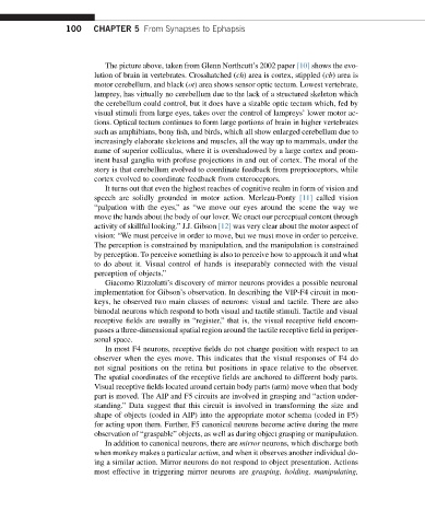
The picture above, taken from Glenn Northcutt’s 2002 paper [10] shows the evo-
lution of brain in vertebrates. Crosshatched (ch) area is cortex, stippled (cb) area is
motor cerebellum, and black (ot) area shows sensor optic tectum. Lowest vertebrate,
lamprey, has virtually no cerebellum due to the lack of a structured skeleton which
the cerebellum could control, but it does have a sizable optic tectum which, fed by
visual stimuli from large eyes, takes over the control of lampreys’ lower motor ac-
tions. Optical tectum continues to form large portions of brain in higher vertebrates
such as amphibians, bony fish, and birds, which all show enlarged cerebellum due to
increasingly elaborate skeletons and muscles, all the way up to mammals, under the
name of superior colliculus, where it is overshadowed by a large cortex and prom-
inent basal ganglia with profuse projections in and out of cortex. The moral of the
story is that cerebellum evolved to coordinate feedback from proprioceptors, while
cortex evolved to coordinate feedback from exteroceptors.
It turns out that even the highest reaches of cognitive realm in form of vision and
speech are solidly grounded in motor action. Merleau-Ponty [11] called vision
“palpation with the eyes,” as “we move our eyes around the scene the way we
move the hands about the body of our lover. We enact our perceptual content through
activity of skillful looking.” J.J. Gibson [12] was very clear about the motor aspect of
vision: “We must perceive in order to move, but we must move in order to perceive.
The perception is constrained by manipulation, and the manipulation is constrained
by perception. To perceive something is also to perceive how to approach it and what
to do about it. Visual control of hands is inseparably connected with the visual
perception of objects.”
Giacomo Rizzolatti’s discovery of mirror neurons provides a possible neuronal
implementation for Gibson’s observation. In describing the VIP-F4 circuit in mon-
keys, he observed two main classes of neurons: visual and tactile. There are also
bimodal neurons which respond to both visual and tactile stimuli. Tactile and visual
receptive fields are usually in “register,” that is, the visual receptive field encom-
passes a three-dimensional spatial region around the tactile receptive field in periper-
sonal space.
In most F4 neurons, receptive fields do not change position with respect to an
observer when the eyes move. This indicates that the visual responses of F4 do
not signal positions on the retina but positions in space relative to the observer.
The spatial coordinates of the receptive fields are anchored to different body parts.
Visual receptive fields located around certain body parts (arm) move when that body
part is moved. The AIP and F5 circuits are involved in grasping and “action under-
standing.” Data suggest that this circuit is involved in transforming the size and
shape of objects (coded in AIP) into the appropriate motor schema (coded in F5)
for acting upon them. Further, F5 canonical neurons become active during the mere
observation of “graspable” objects, as well as during object grasping or manipulation.
In addition to canonical neurons, there are mirror neurons, which discharge both
when monkey makes a particular action, and when it observes another individual do-
ing a similar action. Mirror neurons do not respond to object presentation. Actions
most effective in triggering mirror neurons are grasping, holding, manipulating,