
Page 35 - Bio Engineering Approaches to Cancer Diagnosis and Treatment
P. 35
32 CHAPTER 2 Diagnostic imaging in cancer
• No radiation
• Can be applied for real-time analysis
• No size limitation
• Not require general anesthesia
The disadvantages of using ultrasound imaging are:
• Unsuitable for imaging lungs and bowel
• Resolution is not as good as CT or MRI
• Image analysis is not easily automated
• No whole-body information
• Target-specific imaging is limited to intravascular compartment
2.4 Nonionizing electromagnetic imaging
Nonionizing electromagnetic radiation refers to the low photon energy portion of the
15
electromagnetic spectrum, from 1 to 3 × 10 Hz. In comparison to ionizing radia-
tion, which the interaction with atom result in remove electron, interaction of nonion-
izing electron leads to an excitation and heat production.
The penetration depth in human body, sites of absorption, and the consequent
side effects of the nonionizing electromagnetic radiation are very much depending to
the particular wavelength.
2.4.1 Kinds of nonionizing electromagnetic imaging
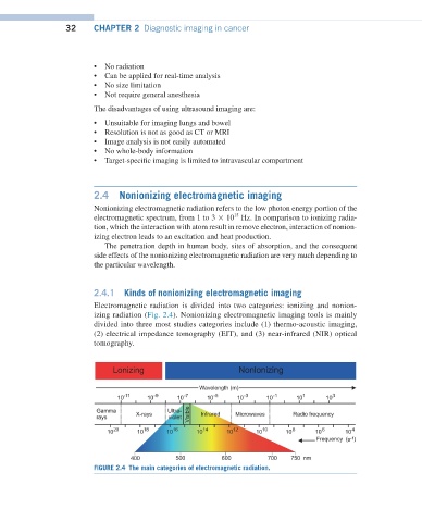
Electromagnetic radiation is divided into two categories: ionizing and nonion-
izing radiation (Fig. 2.4). Nonionizing electromagnetic imaging tools is mainly
divided into three most studies categories include (1) thermo-acoustic imaging,
(2) electrical impedance tomography (EIT), and (3) near-infrared (NIR) optical
tomography.
FIGURE 2.4 The main categories of electromagnetic radiation.