
Page 30 - Bio Engineering Approaches to Cancer Diagnosis and Treatment
P. 30
2.2 Magnetic resonance systems 27
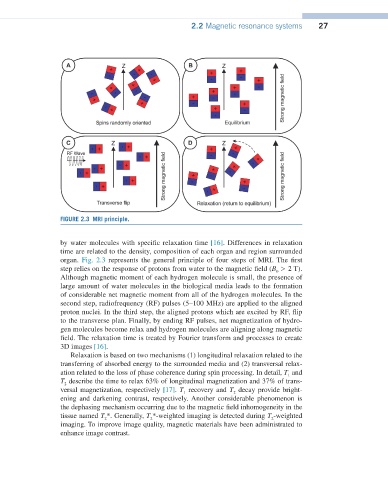
FIGURE 2.3 MRI principle.
by water molecules with specific relaxation time [16]. Differences in relaxation
time are related to the density, composition of each organ and region surrounded
organ. Fig. 2.3 represents the general principle of four steps of MRI. The first
step relies on the response of protons from water to the magnetic field (B > 2 T).
0
Although magnetic moment of each hydrogen molecule is small, the presence of
large amount of water molecules in the biological media leads to the formation
of considerable net magnetic moment from all of the hydrogen molecules. In the
second step, radiofrequency (RF) pulses (5–100 MHz) are applied to the aligned
proton nuclei. In the third step, the aligned protons which are excited by RF, flip
to the transverse plan. Finally, by ending RF pulses, net magnetization of hydro-
gen molecules become relax and hydrogen molecules are aligning along magnetic
field. The relaxation time is treated by Fourier transform and processes to create
3D images [16].
Relaxation is based on two mechanisms (1) longitudinal relaxation related to the
transferring of absorbed energy to the surrounded media and (2) transversal relax-
ation related to the loss of phase coherence during spin processing. In detail, T and
1
T describe the time to relax 63% of longitudinal magnetization and 37% of trans-
2
versal magnetization, respectively [17]. T recovery and T decay provide bright-
2
1
ening and darkening contrast, respectively. Another considerable phenomenon is
the dephasing mechanism occurring due to the magnetic field inhomogeneity in the
tissue named T *. Generally, T *-weighted imaging is detected during T -weighted
2
2
2
imaging. To improve image quality, magnetic materials have been administrated to
enhance image contrast.