
Page 28 - Bio Engineering Approaches to Cancer Diagnosis and Treatment
P. 28
2.1 X-ray-based systems including CT scan 25
a value between +400 and +1000 HU. HU value is determined according to the fol-
lowing equation [3]:
µ µ
−
HU = 1000 × water (2.1)
µ water HU=1000×µ−µwaterµwater
where µ and µ water are the X-ray attenuation coefficient of the desired target and
water, respectively.
One of the main limitations of this technic is related to the low difference between
HU value of soft tissues making the imaging process difficult. To tackle the problem,
radiopaque contrast agents (CAs) are introduced which improve the sensitivity of
X-ray scanner.
2.1.2 Kinds of radiopaque contrast agent
To increase the accuracy of image obtained by X-ray, radiopaque CAs are admin-
istrated during imaging process. By increasing the amount of radiopaque CAs, the
contrast of image is increased in a desired region.
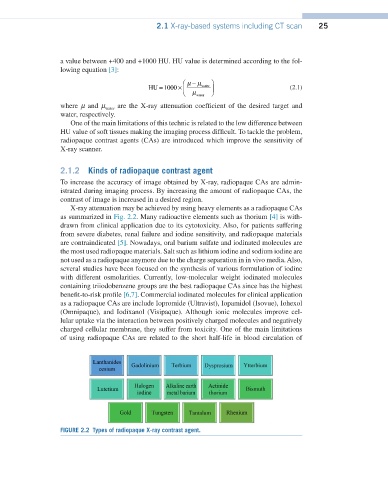
X-ray attenuation may be achieved by using heavy elements as a radiopaque CAs
as summarized in Fig. 2.2. Many radioactive elements such as thorium [4] is with-
drawn from clinical application due to its cytotoxicity. Also, for patients suffering
from severe diabetes, renal failure and iodine sensitivity, and radiopaque materials
are contraindicated [5]. Nowadays, oral barium sulfate and iodinated molecules are
the most used radiopaque materials. Salt such as lithium iodine and sodium iodine are
not used as a radiopaque anymore due to the charge separation in in vivo media. Also,
several studies have been focused on the synthesis of various formulation of iodine
with different osmolarities. Currently, low-molecular weight iodinated molecules
containing triiodobenzene groups are the best radiopaque CAs since has the highest
benefit-to-risk profile [6,7]. Commercial iodinated molecules for clinical application
as a radiopaque CAs are include Iopromide (Ultravist), Iopamidol (Isovue), Iohexol
(Omnipaque), and Iodixanol (Visipaque). Although ionic molecules improve cel-
lular uptake via the interaction between positively charged molecules and negatively
charged cellular membrane, they suffer from toxicity. One of the main limitations
of using radiopaque CAs are related to the short half-life in blood circulation of
FIGURE 2.2 Types of radiopaque X-ray contrast agent.