
Page 190 - Biodegradable Polyesters
P. 190
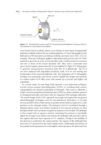
168  7 Electrospun Scaffolds of Biodegradable Polyesters: Manufacturing and Biomedical Application

                                               Gas out
                                Gas in


                                                       hν

                    Polymer sample

                                                   Quartz window

                    Figure 7.3 Photochemical reactor used for UV-assisted functionalization of polymer films in
                    the presence of a gas-reactive atmosphere.

                    cross-linked hybrid scaffolds, photo-cross-linking of electrospun biodegradable
                    polyester scaffolds without the use of photoinitiators, UV laser lithography for the
                    fabrication of 3D microstructured tissue scaffolds, and many more [108–125]. For
                    example, Chen and colleagues applied a microfabrication process, the UV-LIGA
                    method, to generate an array of microneedles with a similar projective structure
                    and size as those of the mouse duodenal villi. They used a commonly used
                    epoxy-based negative photoresist (SU-8) and applied UV-light (UV) lithography
                    to generate microprojective structures from the SU-8 photoresist. The SU-8
                    was then replaced with degradable poly(lactic acid), PLA for cell seeding and
                    proliferation of the intestinal epithelial cells. The integration of UV lithography,
                    molding, hot embossing, and solvent erosion enabled the design and synthesis
                    of a tissue-culture PLA villus array with smooth tip curvature and higher cell
                    population.
                      In a recent study, Gu and Tang [126] reported a new patterning technique,
                    termed enzyme-assisted photolithography (EAPL), to simultaneously achieve
                    topographical and chemical patterning of hydrogels. They used as substrate a
                    widely used biocompatible polyethylene glycol (PEG) to obtain arbitrary patterns
                    of biological molecules and intact cells on integrated PEG hydrogel substrates.
                    The overall design of the EAPL approach was simple and inspired by the general
                    photolithography techniques used for microelectronic fabrication. A two-step
                    process parallel to that of fabricating via positive photoresist is employed to create
                    patterns on the hydrogel surface. The hydrogel is first UV-irradiated through a
                    designed photo mask; cross-linkers localized at the exposed portions are then
                    decaged and the native protease recognition sequences are revealed. The hydrogel
                    is then treated with an aqueous solution containing the protease to specifically
                    digest the decaged cross-linkers and remove the hydrogel with precision only in
                    the regions that have been exposed to UV radiation. Owing to the inability of
                    the protease to proteolyze the caged peptides, the regions of the hydrogels that
                    have been UV protected by the mask remain intact. Additionally, proteolysis of
                    the peptide bonds generates free nucleophilic amine groups at the patterned area