
Page 313 - Biodegradable Polyesters
P. 313
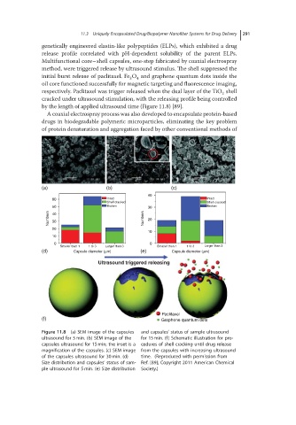
11.3  Uniquely Encapsulated Drug/Biopolymer Nanofiber Systems for Drug Delivery  291

               genetically engineered elastin-like polypeptides (ELPs), which exhibited a drug
               release profile correlated with pH-dependent solubility of the parent ELPs.
               Multifunctional core–shell capsules, one-step fabricated by coaxial electrospray
               method, were triggered release by ultrasound stimulus. The shell suppressed the
               initial burst release of paclitaxel. Fe O and graphene quantum dots inside the
                                           3
                                             4
               oil core functioned successfully for magnetic targeting and fluorescence imaging,
               respectively. Paclitaxel was trigger released when the dual layer of the TiO shell
                                                                         2
               cracked under ultrasound stimulation, with the releasing profile being controlled
               by the length of applied ultrasound time (Figure 11.8) [89].
                A coaxial electrospray process was also developed to encapsulate protein-based
               drugs in biodegradable polymeric microparticles, eliminating the key problem
               of protein denaturation and aggregation faced by other conventional methods of


               (a)                  (b)                  (c)
                                                 40
                  60                Intact                          Intact
                                    Shell cracked                   Shell cracked
                  50                Broken       30                 Broken
                 Numbers  40                    Numbers  20
                  30
                  20
                                                 10
                  10
                   0                              0
                     Smaller than 1  1 to 3  Larger than 3  Smaller than 1  1 to 3  Larger than 3
               (d)       Capsule diameter (μm)  (e)      Capsule diameter (μm)
                                 Ultrasound triggered releasing


                                                      Paclitaxel
               (f)                                    Graphene quantum dots
               Figure 11.8 (a) SEM image of the capsules  and capsules’ status of sample ultrasound
               ultrasound for 5 min. (b) SEM image of the  for 15 min. (f) Schematic illustration for pro-
               capsules ultrasound for 15 min; the inset is a  cedures of shell cracking until drug release
               magnification of the capsules. (c) SEM image  from the capsules with increasing ultrasound
               of the capsules ultrasound for 30 min. (d)  time. (Reproduced with permission from
               Size distribution and capsules’ status of sam-  Ref. [89]; Copyright 2011 American Chemical
               ple ultrasound for 5 min. (e) Size distribution  Society.)