
Page 310 - Biodegradable Polyesters
P. 310
288 11 Electrospun Biopolymer Nanofibers and Their Composites for Drug Delivery Applications
(a) (b)
(c) (d)
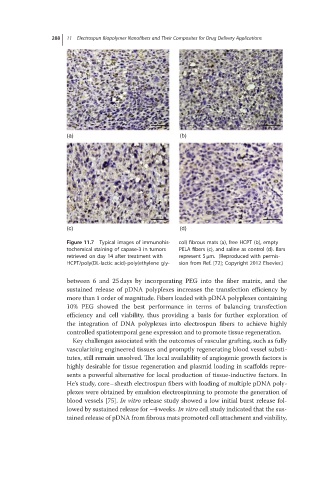
Figure 11.7 Typical images of immunohis- col) fibrous mats (a), free HCPT (b), empty
tochemical staining of capase-3 in tumors PELA fibers (c), and saline as control (d). Bars
retrieved on day 14 after treatment with represent 5 μm. (Reproduced with permis-
HCPT/poly(DL-lactic acid)-poly(ethylene gly- sion from Ref. [72]; Copyright 2012 Elsevier.)
between 6 and 25 days by incorporating PEG into the fiber matrix, and the
sustained release of pDNA polyplexes increases the transfection efficiency by
more than 1 order of magnitude. Fibers loaded with pDNA polyplexes containing
10% PEG showed the best performance in terms of balancing transfection
efficiency and cell viability, thus providing a basis for further exploration of
the integration of DNA polyplexes into electrospun fibers to achieve highly
controlled spatiotemporal gene expression and to promote tissue regeneration.
Key challenges associated with the outcomes of vascular grafting, such as fully
vascularizing engineered tissues and promptly regenerating blood vessel substi-
tutes, still remain unsolved. The local availability of angiogenic growth factors is
highly desirable for tissue regeneration and plasmid loading in scaffolds repre-
sents a powerful alternative for local production of tissue-inductive factors. In
He’s study, core–sheath electrospun fibers with loading of multiple pDNA poly-
plexes were obtained by emulsion electrospinning to promote the generation of
blood vessels [75]. In vitro release study showed a low initial burst release fol-
lowed by sustained release for ∼4weeks. In vitro cell study indicated that the sus-
tained release of pDNA from fibrous mats promoted cell attachment and viability,