
Page 291 - Biomedical Engineering and Design Handbook Volume 2, Applications
P. 291
THE PRINCIPLES OF X-RAY COMPUTED TOMOGRAPHY 269
energy sensitive and provide information of the chemical composition in addition to structural mor-
phology. Further, by optically collimating the primary x-ray beam to a secondary focus, it is possi-
ble to form images by the x-ray fluorescence emission process and hence provide a map of the
atomic composition.
A further indication of the versatility of the projection x-ray microscope is the possibility of splitting
the beam path in various ways to provide enhanced imaging detail by phase contrast. Although the
assumption that diffraction effects may be neglected has proved to be an acceptable approximation for
most attenuation contrast imaging applications, they can nevertheless be manipulated to reveal additional
information. A medium of varying density will, by virtue of the associated changes in refractive index,
8
create phase differences in the propagating waves of adjacent ray-paths. Under normal circumstances
these effects are small and would not be seen in the image. However, if the difference in phase is con-
verted into spatial displacement, very fine detail emerge as phase contrast in the image. This method is
currently receiving a great deal of attention as means of providing improved contrast. 9
10.2 THE INTERACTION OF X-RAYS WITH MATTER
10.2.1 The Collision Model
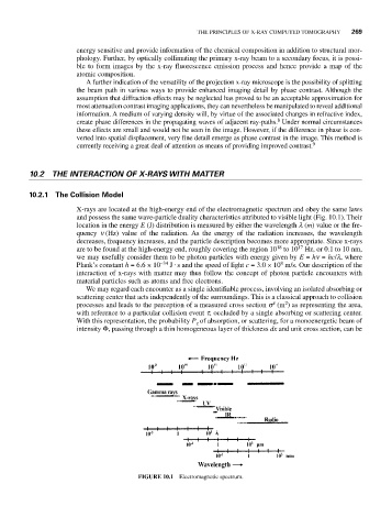
X-rays are located at the high-energy end of the electromagnetic spectrum and obey the same laws
and possess the same wave-particle duality characteristics attributed to visible light (Fig. 10.1). Their
location in the energy E (J) distribution is measured by either the wavelength l (m) value or the fre-
quency n (Hz) value of the radiation. As the energy of the radiation increases, the wavelength
decreases, frequency increases, and the particle description becomes more appropriate. Since x-rays
18
17
are to be found at the high-energy end, roughly covering the region 10 to 10 Hz, or 0.1 to 10 nm,
we may usefully consider them to be photon particles with energy given by E = hn = hc/l, where
8
Plank’s constant h = 6.6 × 10 −34 J ⋅ s and the speed of light c = 3.0 × 10 m/s. Our description of the
interaction of x-rays with matter may thus follow the concept of photon particle encounters with
material particles such as atoms and free electrons.
We may regard each encounter as a single identifiable process, involving an isolated absorbing or
scattering center that acts independently of the surroundings. This is a classical approach to collision
t
2
processes and leads to the perception of a measured cross section s (m ) as representing the area,
with reference to a particular collision event t, occluded by a single absorbing or scattering center.
With this representation, the probability P of absorption, or scattering, for a monoenergetic beam of
t
intensity Φ, passing through a thin homogeneous layer of thickness dx and unit cross section, can be
FIGURE 10.1 Electromagnetic spectrum.